10 The Sensory Systems
In this laboratory session, we will study aspects of the anatomy of the visual, olfactory, gustatory, and auditory systems. Below, you will be presented with a number of figures and asked to label or color certain structures in each figure.
10.1 The Visual System
10.1.1 Eyball Dissection
10.1.2 The Eye
In Figure 10.1 , label the following structures:
- Color the lens of the eye in yellow.
- Color the vitreous body in pink.
- Color the iris in brown.
- Color the retina in green.
- Color the sclera in blue.
- Color the ciliary body in red.
- Label the structures pointed to by the black lines with the appropriate anatomical name.
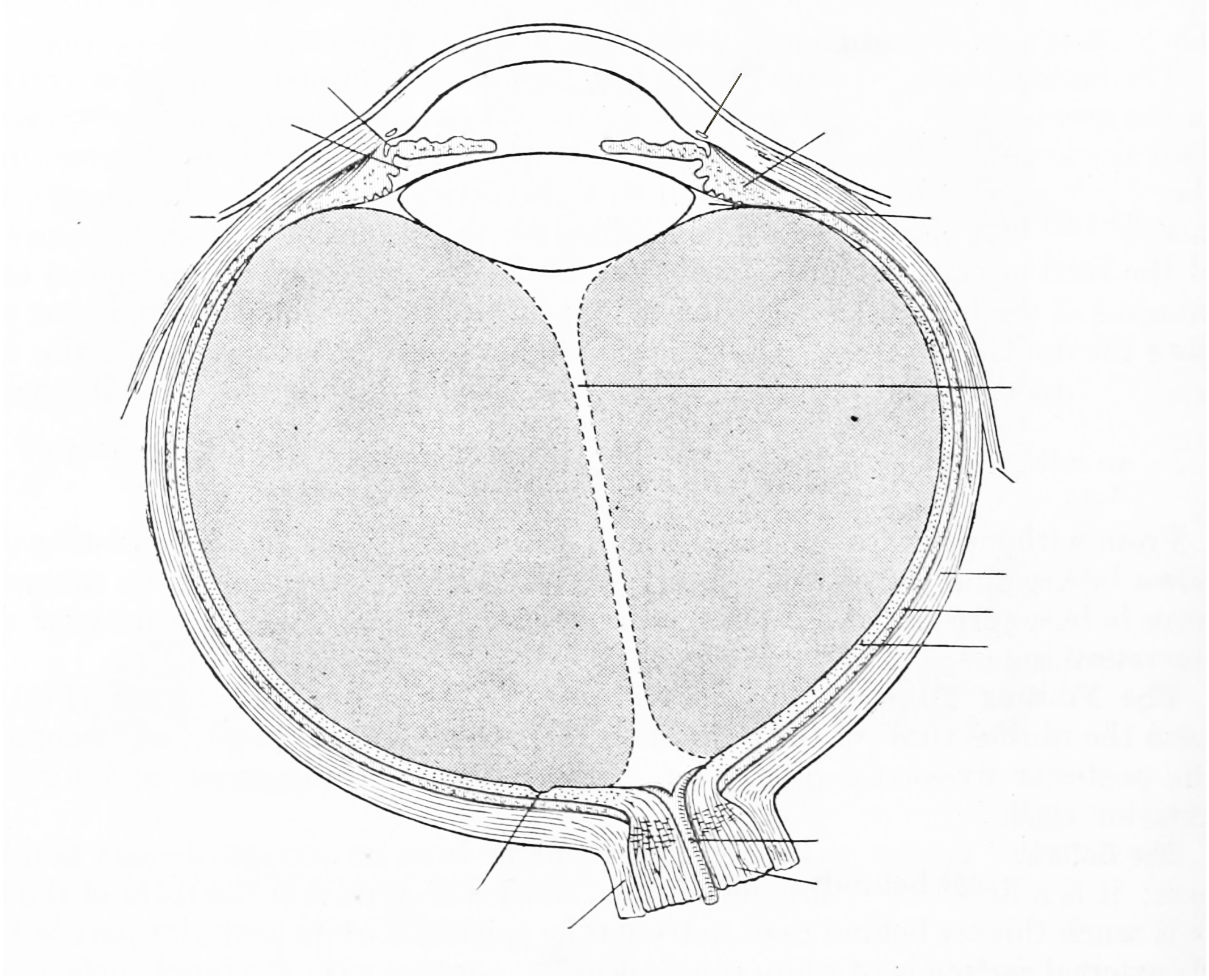
Figure 10.1: A horizontal section of the eyball.
10.1.3 The Retina
In Figure 10.2 , label the following layers, structures, and cells:
- Photoreceptor layer.
- Outer plexiform layer.
- Internal nuclear layer.
- Internal plexiform layer.
- Ganglion cell layer.
- Ganglion cell axons.
- A cone.
- A rod.
- Horizontal cells.
- Amacrine cells.
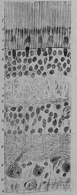
Figure 10.2: Vertical section of the adult human retina. Carmine and Nissl stain. Modified from Fig. 188 in Histologie du système nerveux de l’homme & des vertébrés (1909) by Santiago Ramón y Cajal translated from Spanish by Dr. L. Azoulay.
In Figure 10.3, label the following layers, structures, and cells:
- A rod
- A cone
- Ahorizontal cell
- A bipolar cell
- Various types of amacrine cells
- A ganglion cell
- A displaced amacrine cell
- A pigment epithelial cell with extended process
- A pigment epithelial cell with retracted process
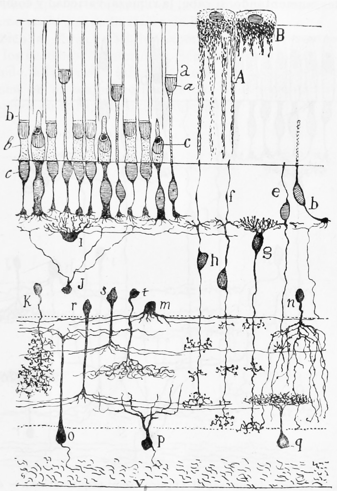
Figure 10.3: A semischematic diagram of the frog retina.
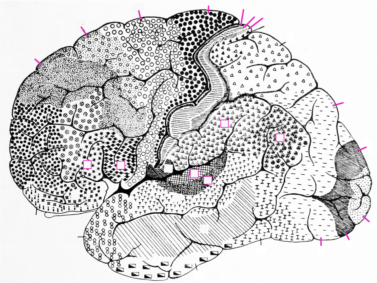
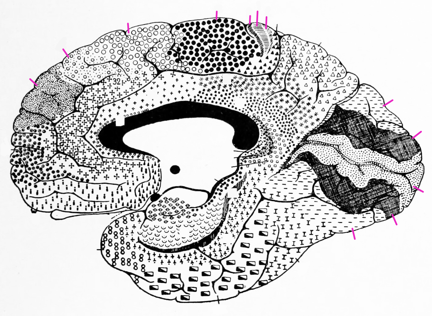
10.1.4 Visual Areas Of The Cortex
10.2 The Olfactory System
In Figure 10.4 , label the following layers, structures, and cells:
- Label the olfacory mucosa.
- Label the glomeruli in the olfactory bulb.
- Label the mitral cells.
- Label the granule cells
- Label the olfactory nerve.
- Label the pyramidal cells in the olfactory cortex.
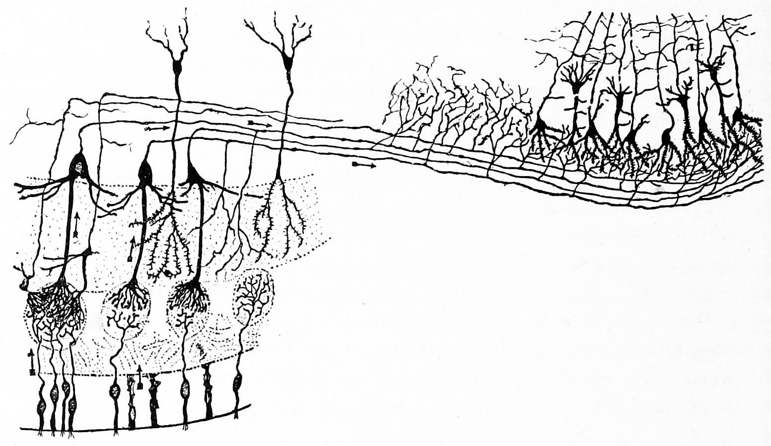
Figure 10.4: Diagram of the structure of the olfactory bulb and olfactory cortex. Histologie du système nerveux de l’homme & des vertébrés, Tome Premier (1909) by Santiago Ramón y Cajal translated from Spanish by Dr. L. Azoulay.
10.3 The Gustatory System
In Figure 10.5 , label the following structures:
- Label a gustatory cell.
- Label a supporting cell.
- Label gustatory hairs.
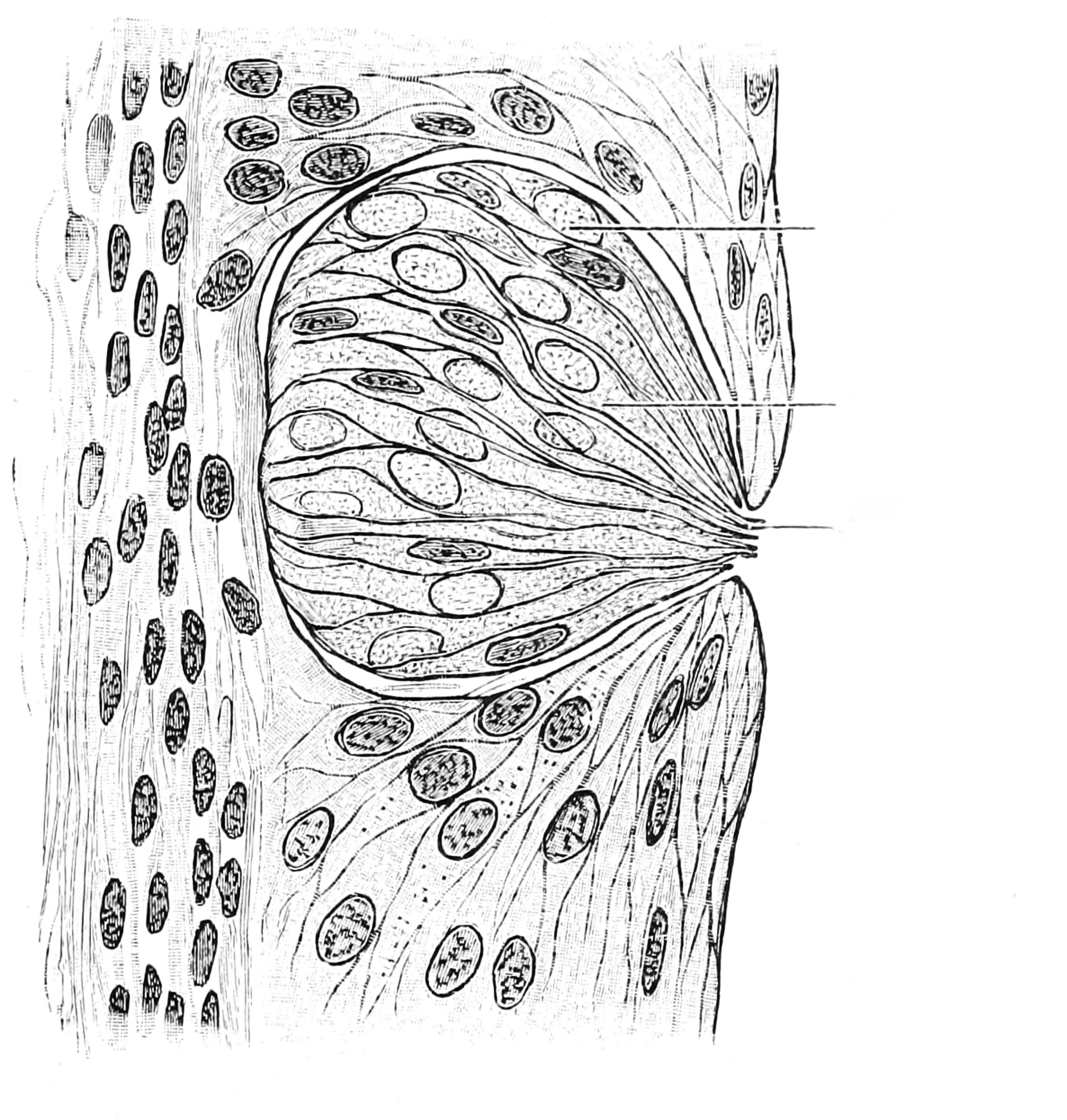
Figure 10.5: Diagram of the structure of the olfactory bulb and olfactory cortex. Histologie du système nerveux de l’homme & des vertébrés, Tome Premier (1909) by Santiago Ramón y Cajal translated from Spanish by Dr. L. Azoulay.
10.4 The Auditory System
In Figure 10.6 , label the following structures:
- Label the Eustachian tube.
- Label the cochlea.
- Label the membrana tympani.
- Label the pinna.
- Label the tympanic cavity with the chain of ossicles (malleus, incus, and stapes).
- Label the sacule.
- Label the utricule.
- Label the semicircular canals.
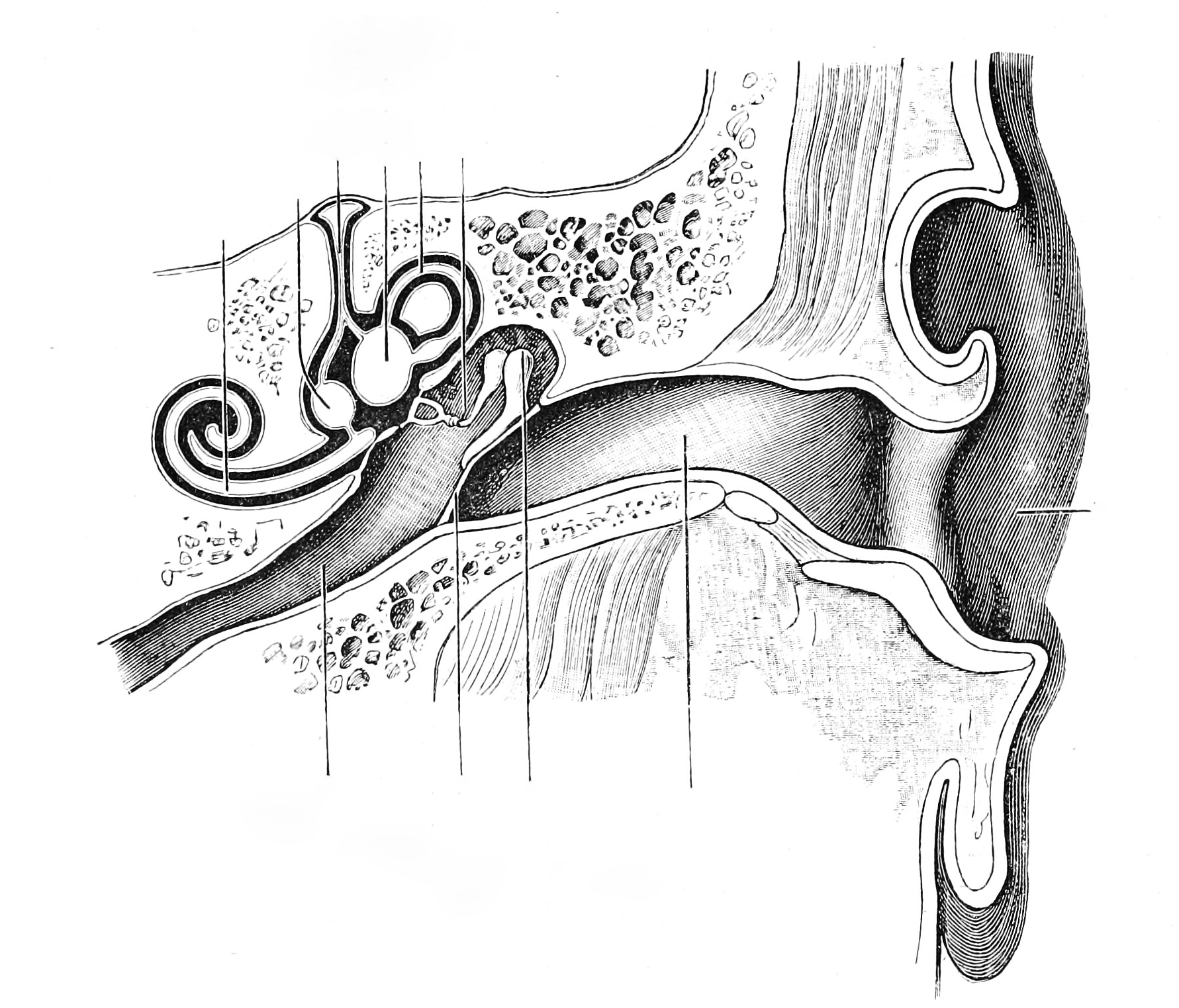
Figure 10.6: Diagrammatic view of the hearing organ. From Textbook of anatomy. Section 2. The muscular system: the nervous system: the organs of sense and integument edited by D. J. Cunningham
In Figure 10.7 , label the following structures:
- Label the vestibular membrane.
- Label the scala vestibuli.
- Label the scala tympani.
- Label the cochlear nerve.
- Label the spiral ganglion.
- Label the organ of Corti.
- Label the basilar membrane.
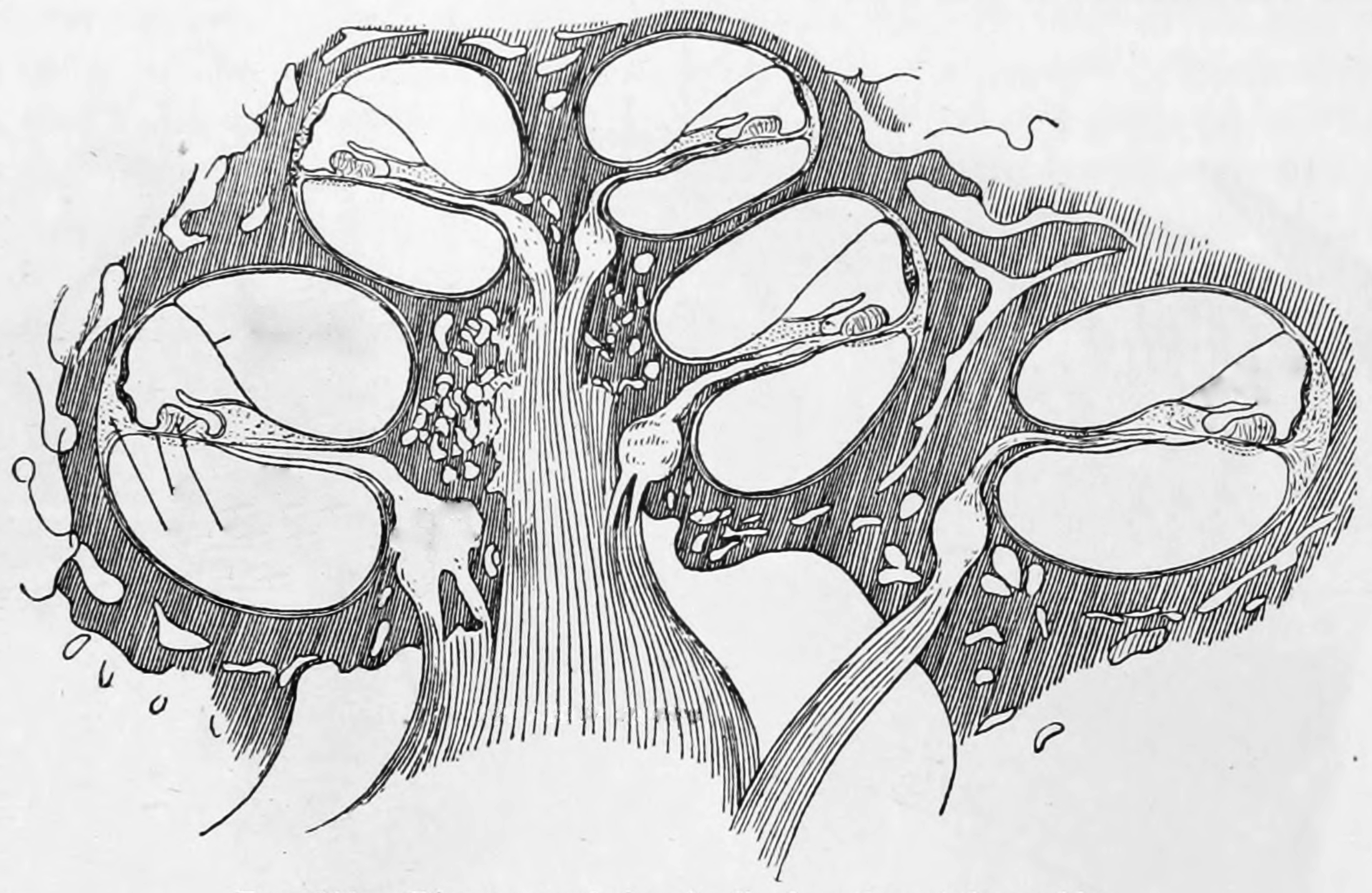
Figure 10.7: Diagramatic longitudinal section of the cochlea. From Gray Henry, Anatomy of the Human Body. 20th Edition, Lea & Febiger, Philadelphia & New York, 1918
In Figure 10.8 , label the following structures:
- Label the membrana tectoria.
- Label the scala outer hair cells.
- Label the scala inner hair cells.
- Label the scala tympani.
- Label the basilar membrane.
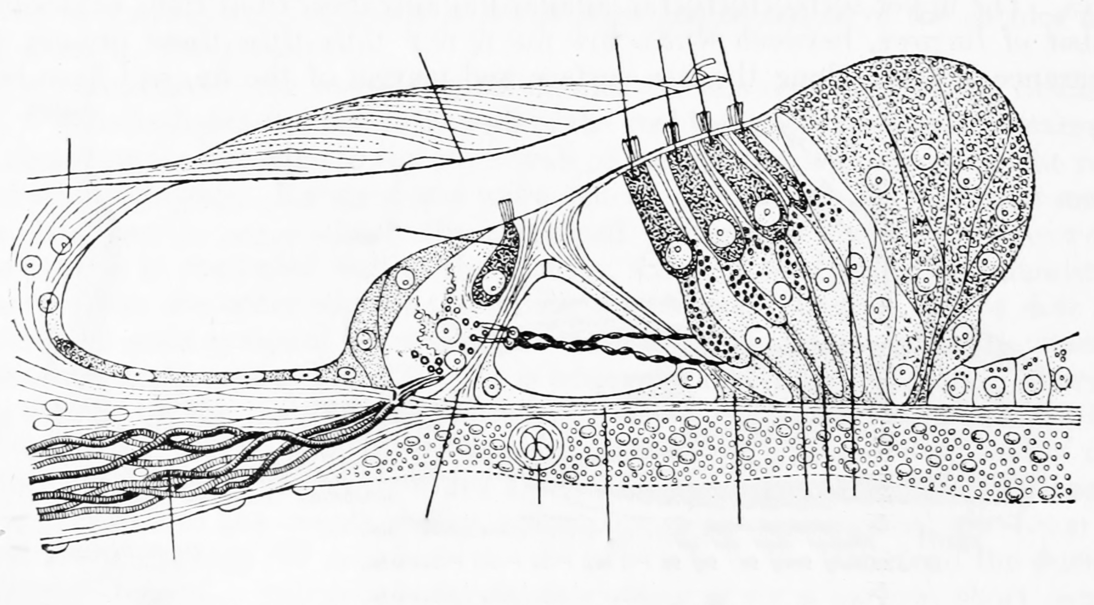
Figure 10.8: Section through the spiral organ of Corti (magnified). From Gray Henry, Anatomy of the Human Body. 20th Edition, Lea & Febiger, Philadelphia & New York, 1918