2 Neurons And Glia
In this laboratory session, we will study the morphology of the basic cellular constitutents of the nervous system: neurons and glia.
Below, you will be presented with a number of figures and asked to label or color certain structures in each figure.
In Figure 2.1 label the following structures
- Dendrites
- Axon hillock
- Nodes of Ranvier
- Myelin sheath
- Neuronal cell membrane
- Golgi apparatus
- Ribosomes
- Rough endoplasmic reticulum
- Smooth endoplasmic reticulum
- Cell nucleus
- Nucleolus
- Mitochondrion
- Microtubules
- Synapse
- Synaptic vesicles
- Synaptic cleft
- Neurotransmitter receptors
- Axon terminal

Figure 2.1: Diagram of a myelinated vertebrate motor neuron.
In Figure 2.2 label the following types of neurons
- Unipolar neurons
- Bipolar neurons
- Golgi I neurons: a Purkinje cell, spinal motor neuron and a pyramidal cell
- Golgi II neuron
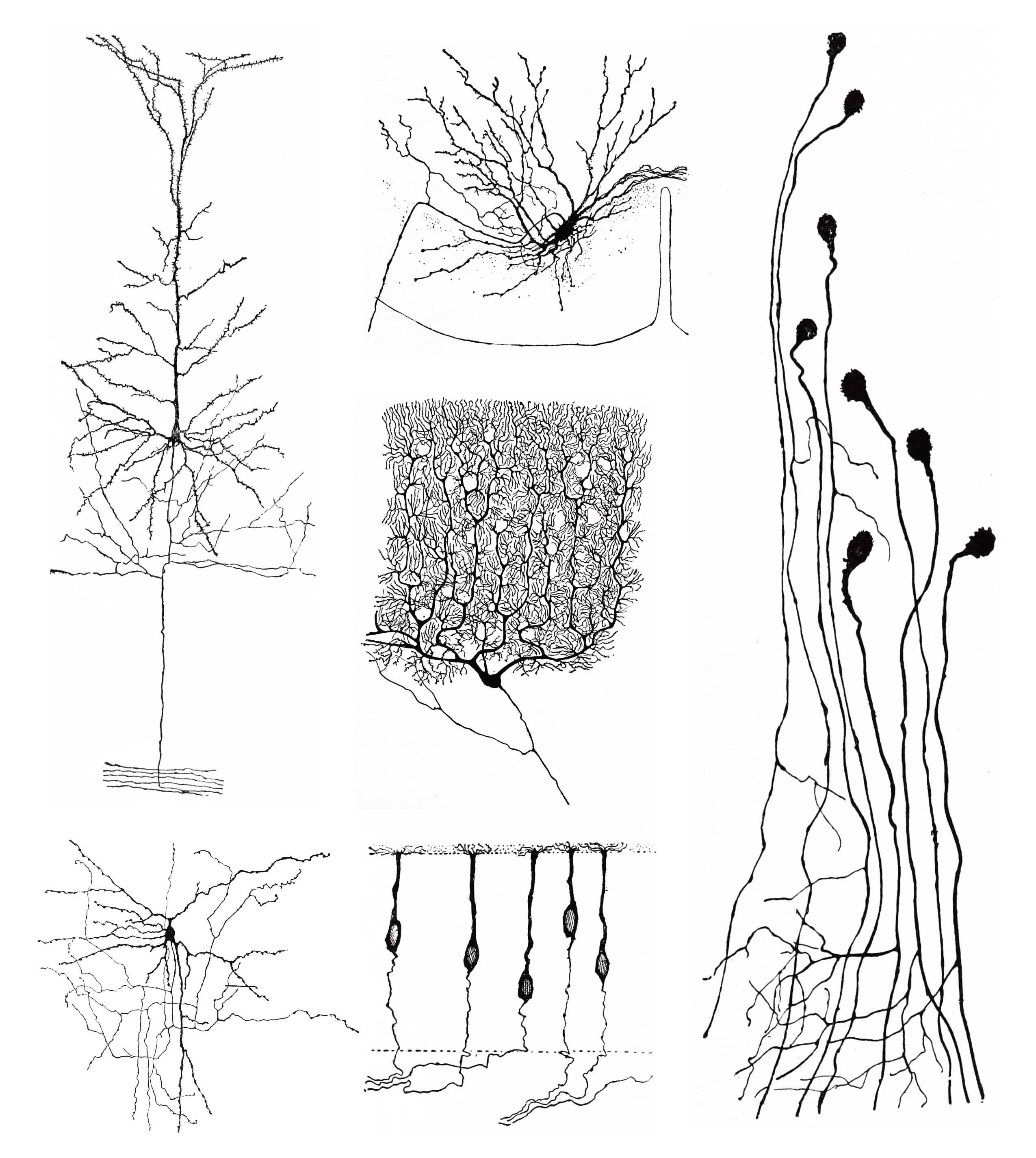
Figure 2.2: Morpholoigcally distinct types of neurons after Cajal. Histologie du système nerveux de l’homme & des vertébrés, Tome Premier (1909) by Santiago Ramón y Cajal translated from Spanish by Dr. L. Azoulay.
In Figure 2.2 label the following types of glial cells
- Astrocytes
- Oligodendrocytes
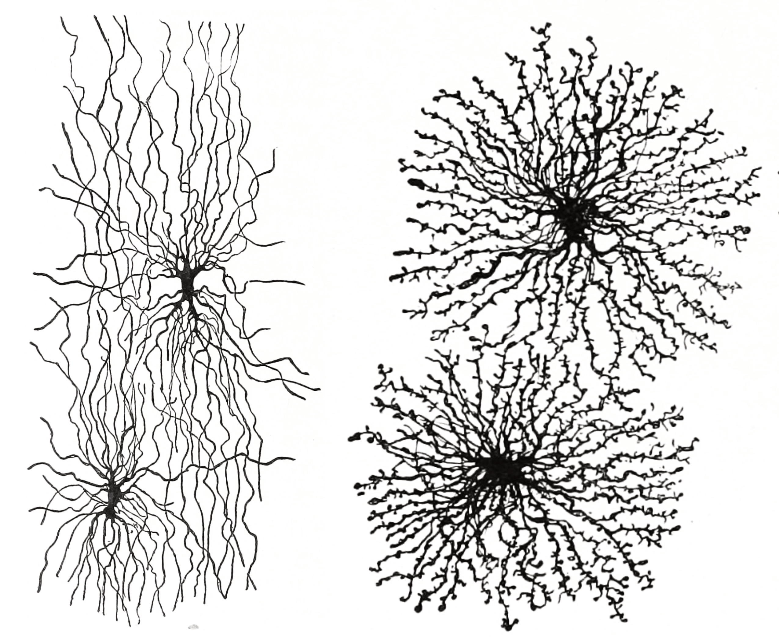
Figure 2.3: Astrocytes and oligodendrocytes are the major types of macroglia in the grey and white matter of the brain, respectively. Histologie du système nerveux de l’homme & des vertébrés, Tome Premier (1909) by Santiago Ramón y Cajal translated from Spanish by Dr. L. Azoulay.