5 The Diencephalon
In this laboratory session, we will study the anatomy of the human diencephalon. The diencephalon is a division of the forebrain (embryonic prosencephalon), and is situated between the telencephalon and the midbrain (embryonic mesencephalon). It consists of structures that are on either side of the third ventricle, including the thalamus, the hypothalamus, the epithalamus and the subthalamus.
Below, you will be presented with a number of figures and asked to label or color certain structures in each figure.
5.1 A Series Of Coronal Sections Of A Human Brain
In Figure 4.1, label the following structures:
- The optic nerves (left and right)
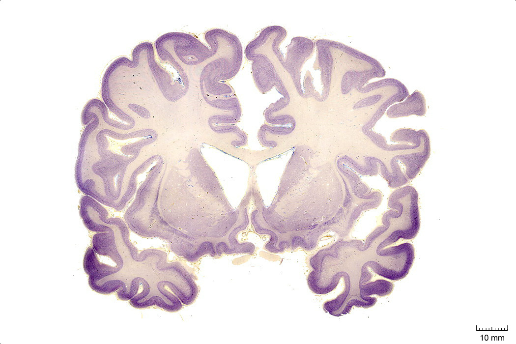
Figure 4.1: Coronal section from The Human Brain Atlas at the Michigan State University Brain Biodiveristy Bank which acknowledges their support from the National Science Foundation.
In Figure 4.2, label the following structures:
- The preoptic area
- The optic chiasm
- The infundibular stalk
- The 3d ventricle
- The internal capsule
- The external capsule
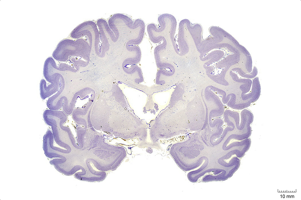
Figure 4.2: Coronal section from The Human Brain Atlas at the Michigan State University Brain Biodiveristy Bank which acknowledges their support from the National Science Foundation.
In Figure 4.3, label the following structures:
- The thalamus
- The hypothalamus
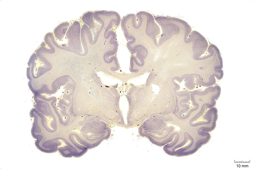
Figure 4.3: Coronal section from The Human Brain Atlas at the Michigan State University Brain Biodiveristy Bank which acknowledges their support from the National Science Foundation.
In Figure 4.4, label the following structures:
- The thalamus
- The 3d ventricle
- The subthalamic nucleus
- The zona incerta
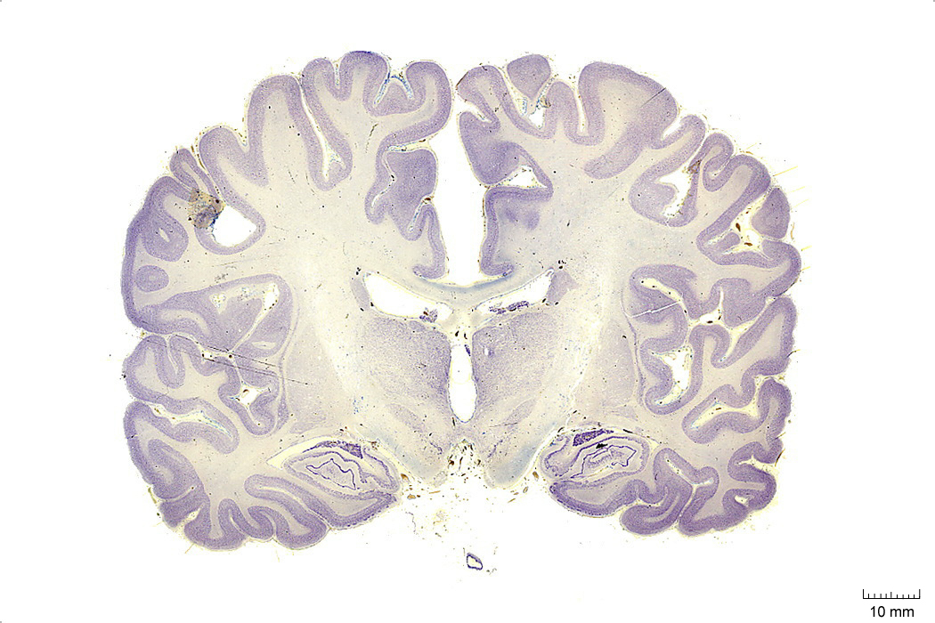
Figure 4.4: Coronal section from The Human Brain Atlas at the Michigan State University Brain Biodiveristy Bank which acknowledges their support from the National Science Foundation.
In Figure 4.5, label the following structures:
- The thalamus
- The 3d ventricle
- The internal capsule
- The external capsule
- The subthalamic nucleus
- The zona incerta
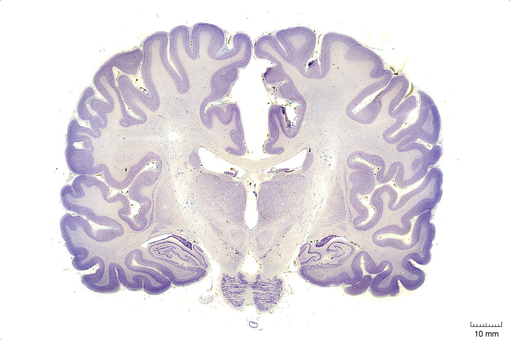
Figure 4.5: Coronal section from The Human Brain Atlas at the Michigan State University Brain Biodiveristy Bank which acknowledges their support from the National Science Foundation.
In Figure 4.6, label the following structures:
- The habenula
- The pineal gland
- The medial geniculate nucleus
- The lateral geniculate nucleus
- The posterior commissure
- The cerebral aqueduct
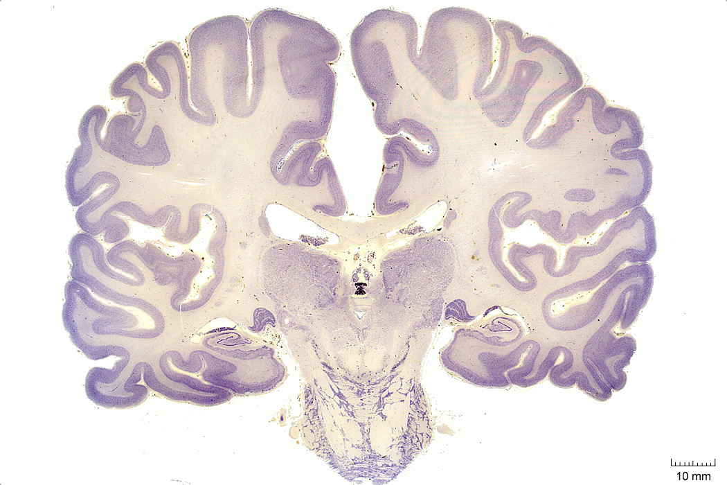
Figure 4.6: Coronal section from The Human Brain Atlas at the Michigan State University Brain Biodiveristy Bank which acknowledges their support from the National Science Foundation.
In Figure 4.7, label the following structures:
- The thalamus
- The pineal gland
- The periaqueductal grey matter
- The cerebral aqueduct
- The pulvinar
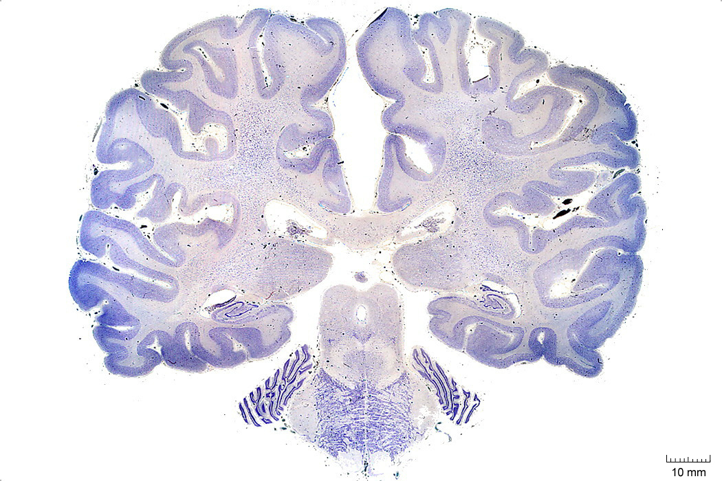
Figure 4.7: Coronal section from The Human Brain Atlas at the Michigan State University Brain Biodiveristy Bank which acknowledges their support from the National Science Foundation.
5.2 A Series Of Sagittal Sections Of A Human Brain
In Figure 4.23, label the following structures:
- The thalamic nuclei
- pulvinar
- ventral group
- centromedian gropup
- The subthalamic nucleus
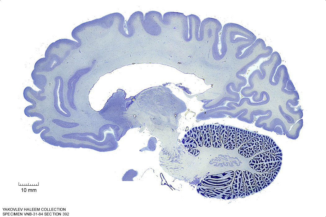
Figure 4.23: Sagittal section from The Human Brain Atlas at the Michigan State University Brain Biodiveristy Bank which acknowledges their support from the National Science Foundation.
In Figure 4.24, label the following structures:
- The thalamic nuclei
- anterior group
- ventral group
- centromedian gropup
- The anterior commissure
- The hypothalamus
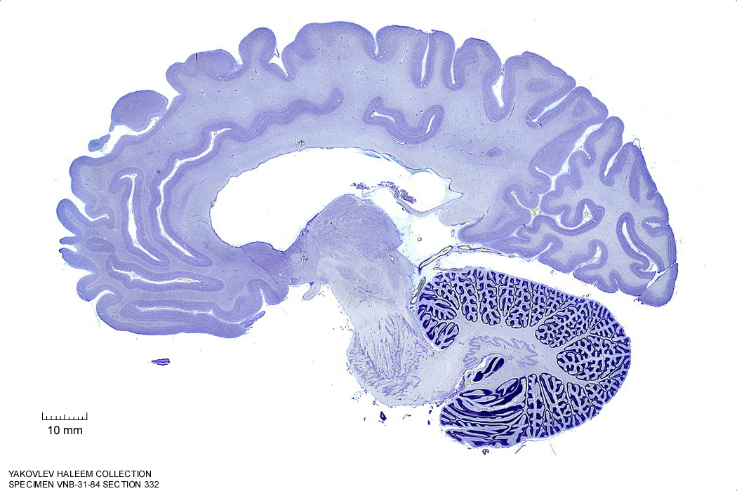
Figure 4.24: Sagittal section from The Human Brain Atlas at the Michigan State University Brain Biodiveristy Bank which acknowledges their support from the National Science Foundation.
In Figure 4.25, label the following structures:
- The thalamus
- The anterior commissure
- The mammillothalamic tract
- The hypothalamus
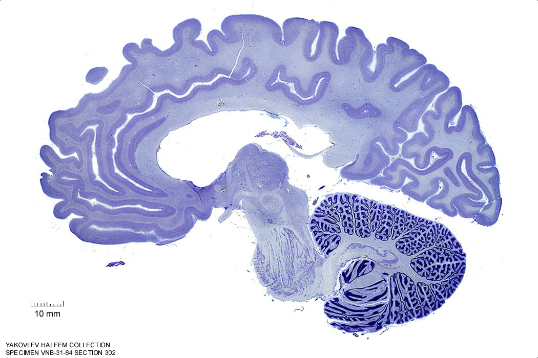
Figure 4.25: Sagittal section from The Human Brain Atlas at the Michigan State University Brain Biodiveristy Bank which acknowledges their support from the National Science Foundation.
In Figure 4.26, label the following structures:
- The thalamus
- The mammillothalamic tract
- The hypothalamus
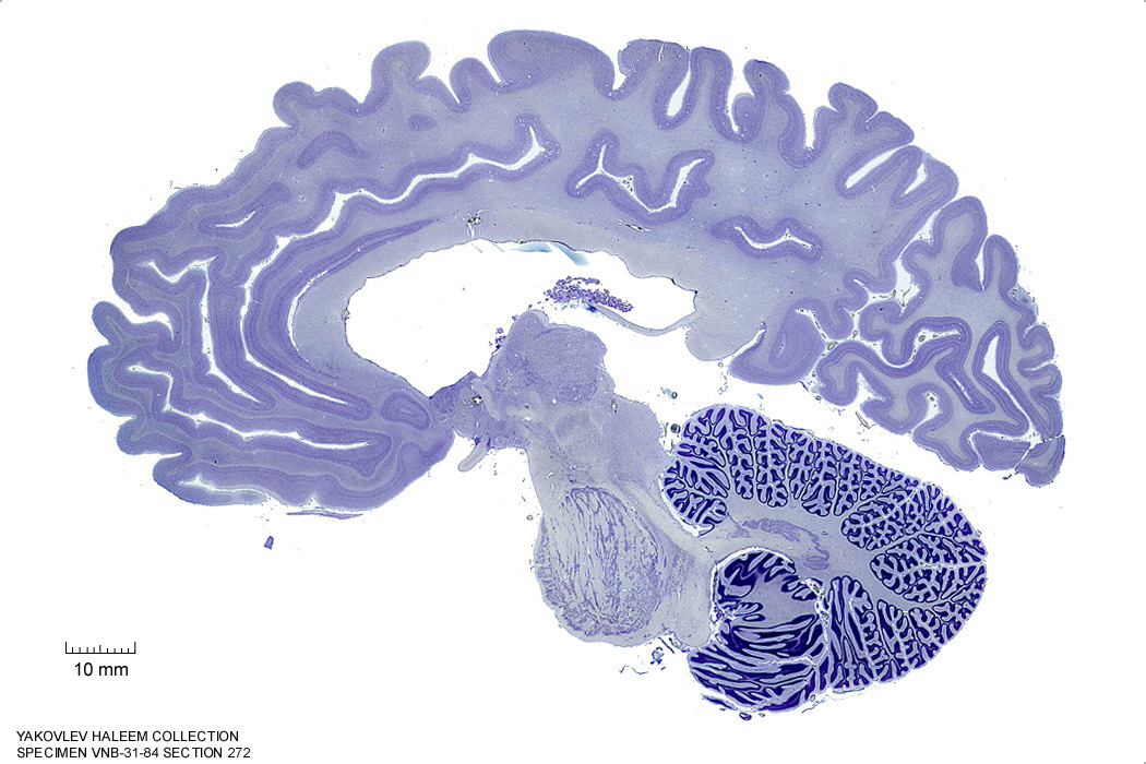
Figure 4.26: Sagittal section from The Human Brain Atlas at the Michigan State University Brain Biodiveristy Bank which acknowledges their support from the National Science Foundation.
In Figure 4.27, label the following structures:
- The thalamus
- The mammillothalamic tract
- The hypothalamus
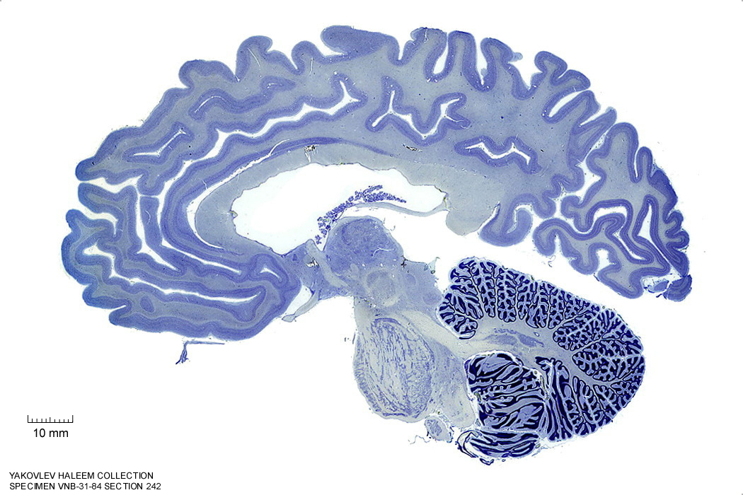
Figure 4.27: Sagittal section from The Human Brain Atlas at the Michigan State University Brain Biodiveristy Bank which acknowledges their support from the National Science Foundation.
In Figure 4.28, label the following structures:
- The thalamus
- The mammillothalamic tract
- The hypothalamus
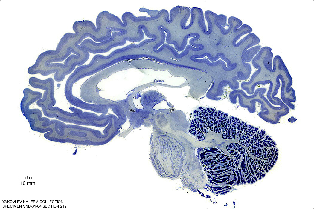
Figure 4.28: Sagittal section from The Human Brain Atlas at the Michigan State University Brain Biodiveristy Bank which acknowledges their support from the National Science Foundation.
In Figure 4.29, label the following structures:
- The thalamus
- The mammillothalamic tract
- The hypothalamus
- The habenular commissure
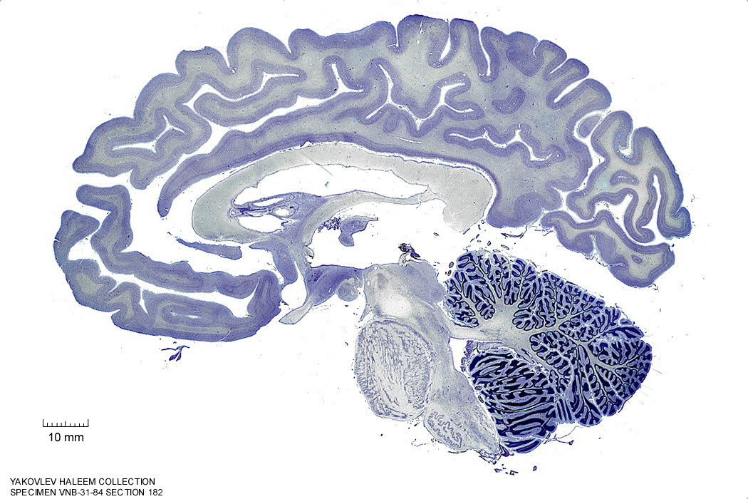
Figure 4.29: Sagittal section from The Human Brain Atlas at the Michigan State University Brain Biodiveristy Bank which acknowledges their support from the National Science Foundation.
In Figure 4.30, label the following structures:
- The thalamus
- The optic chiasm
- The mammillothalamic tract
- The hypothalamus
- The cerebral aqueduct
- The posterior commissure
Figure 4.30: Sagittal section from The Human Brain Atlas at the Michigan State University Brain Biodiveristy Bank which acknowledges their support from the National Science Foundation.
In Figure 5.1, label the structures listed on the bottom left side:
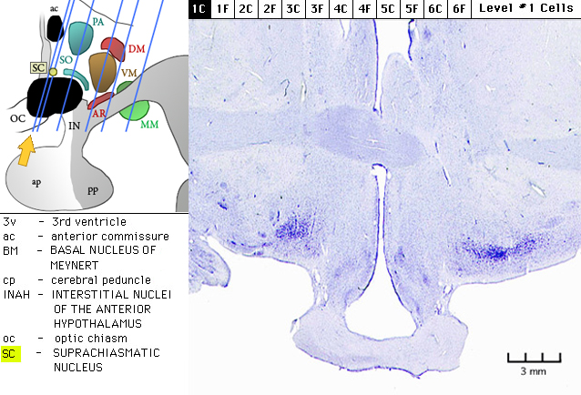
Figure 5.1: Sagittal section from The Human Brain Atlas at the Michigan State University Brain Biodiveristy Bank which acknowledges their support from the National Science Foundation.
In Figure 5.2, label the structures listed on the bottom left side:
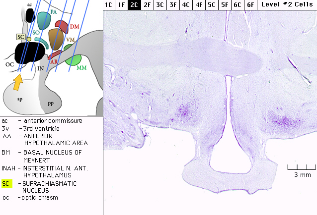
Figure 5.2: Sagittal section from The Human Brain Atlas at the Michigan State University Brain Biodiveristy Bank which acknowledges their support from the National Science Foundation.
In Figure 5.3, label the structures listed on the bottom left side:
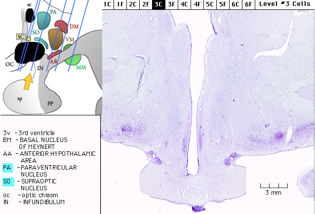
Figure 5.3: Sagittal section from The Human Brain Atlas at the Michigan State University Brain Biodiveristy Bank which acknowledges their support from the National Science Foundation.
In Figure 5.4, label the structures listed on the bottom left side:
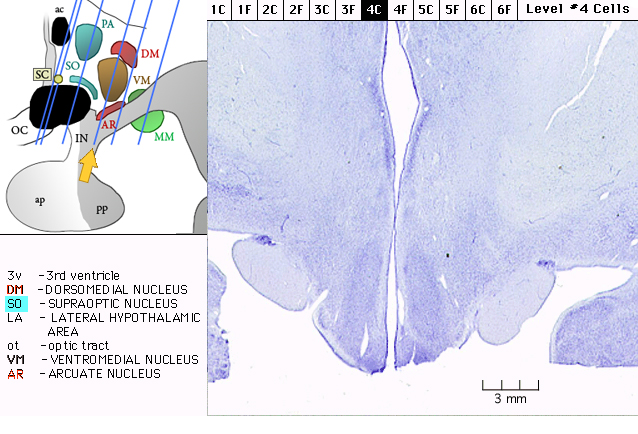
Figure 5.4: Sagittal section from The Human Brain Atlas at the Michigan State University Brain Biodiveristy Bank which acknowledges their support from the National Science Foundation.
In Figure 5.5, label the structures listed on the bottom left side:
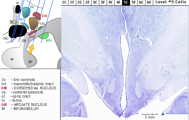
Figure 5.5: Sagittal section from The Human Brain Atlas at the Michigan State University Brain Biodiveristy Bank which acknowledges their support from the National Science Foundation.
In Figure 5.6, label the structures listed on the bottom left side:
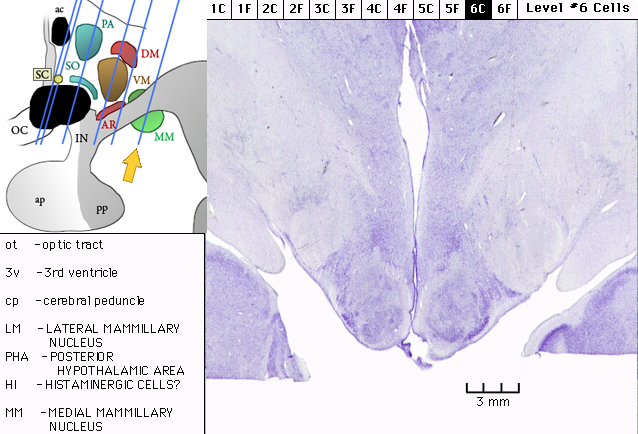
Figure 5.6: Sagittal section from The Human Brain Atlas at the Michigan State University Brain Biodiveristy Bank which acknowledges their support from the National Science Foundation.
5.3 A Series Of Horizontal Sections Of A Human Brain
In Figure 4.36, label the following structures:
- The thalamic nuclei
- reticular
- ventroposterior
- ventroanterior
- ventrolateral
- mediodorsal
- anteroprincipal
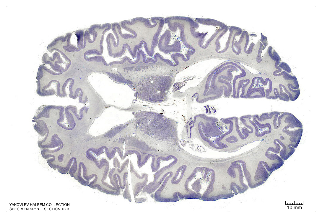
Figure 4.36: Horizontal section from The Human Brain Atlas at the Michigan State University Brain Biodiveristy Bank which acknowledges their support from the National Science Foundation.
In Figure 4.37, label the following structures:
- The thalamic nuclei
- pulvinar
- reticular
- ventroposterior
- ventroanterior
- ventrolateral
- mediodorsal
- The habenula
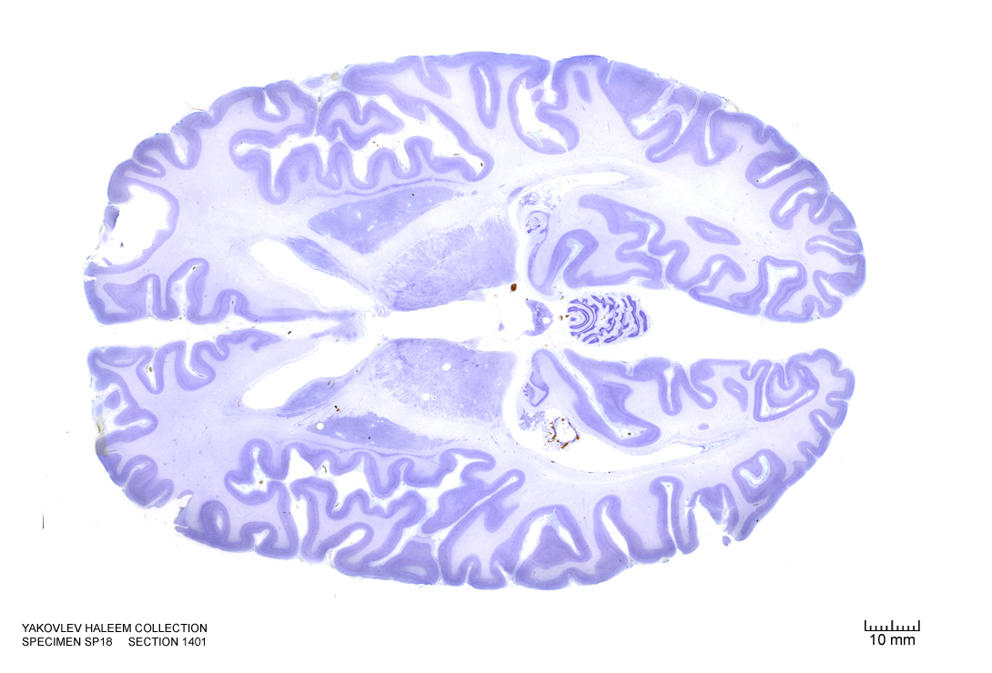
Figure 4.37: Horizontal section from The Human Brain Atlas at the Michigan State University Brain Biodiveristy Bank which acknowledges their support from the National Science Foundation.
In Figure 4.38, label the following structures:
- The thalamic nuclei
- pulvinar
- reticular
- The 3d ventricle
- The medial geniculate nucleus
- The lateral geniculate nucleus
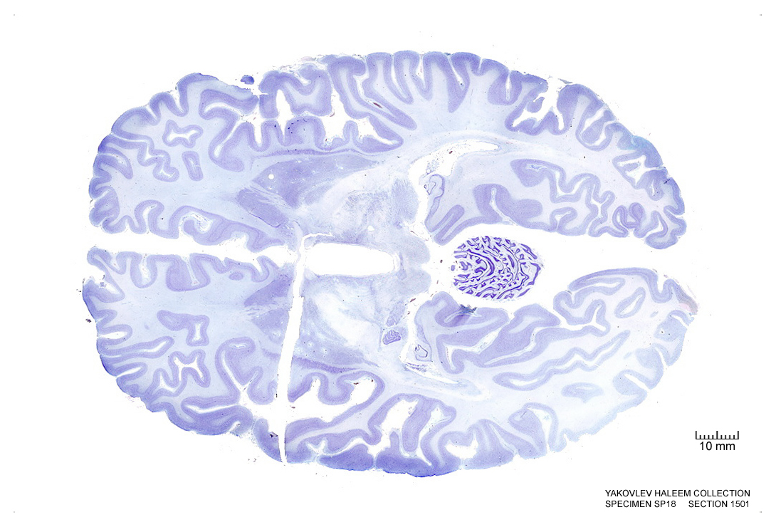
Figure 4.38: Horizontal section from The Human Brain Atlas at the Michigan State University Brain Biodiveristy Bank which acknowledges their support from the National Science Foundation.
In Figure 4.39, label the following structures:
- The cerebral aqueduct
- The mammillothalamic tract
- The medial geniculate nucleus
- The lateral geniculate nucleus
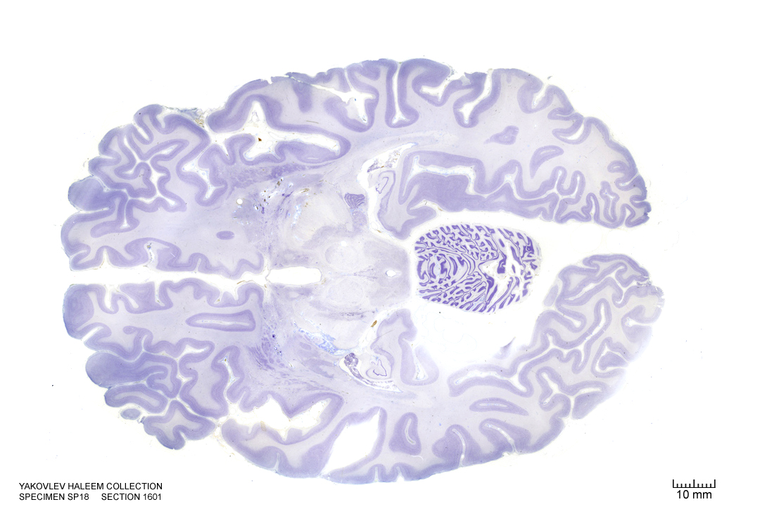
Figure 4.39: Horizontal section from The Human Brain Atlas at the Michigan State University Brain Biodiveristy Bank which acknowledges their support from the National Science Foundation.
In Figure 4.40, label the following structures:
- The cerebral aqueduct
- The periaqueductal grey area
- The 3d ventricle
- The medial geniculate nucleus
- The lateral geniculate nucleus
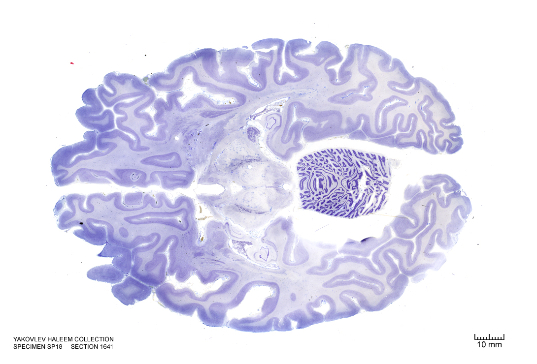
Figure 4.40: Horizontal section from The Human Brain Atlas at the Michigan State University Brain Biodiveristy Bank which acknowledges their support from the National Science Foundation.
In Figure 4.41, label the following structures:
- The cerebral aqueduct
- The periaqueductal grey area
- The 3d ventricle
- The medial geniculate nucleus
- The lateral geniculate nucleus
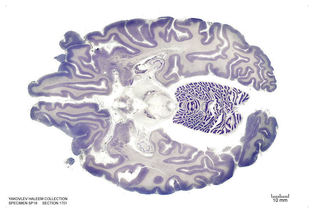
Figure 4.41: Horizontal section from The Human Brain Atlas at the Michigan State University Brain Biodiveristy Bank which acknowledges their support from the National Science Foundation.
In Figure 4.42, label the following structures:
- The cerebral aqueduct
- The arcuate hypothalamic nucleus
- The periaqueductal grey area
- The 3d ventricle
- The 4th ventricle
- The raphe nucleus
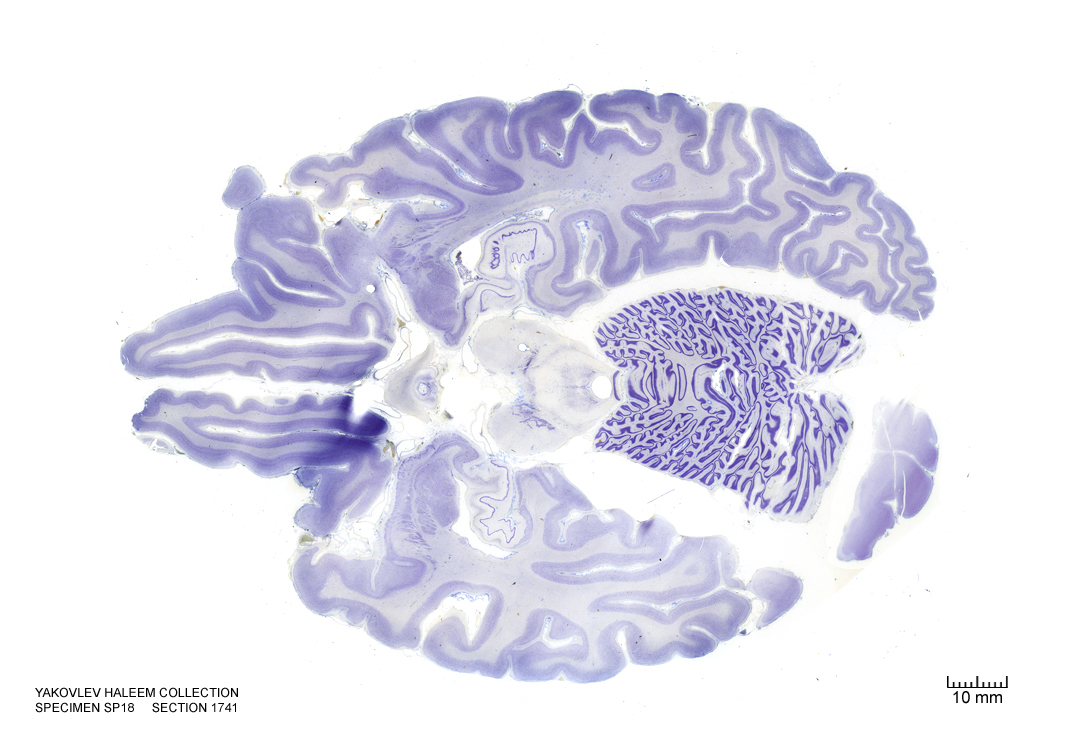
Figure 4.42: Horizontal section from The Human Brain Atlas at the Michigan State University Brain Biodiveristy Bank which acknowledges their support from the National Science Foundation.
In Figure 4.43, label the following structures:
- The arcuate hypothalamic nucleus
- The infundibular stalk
- The raphe nuclei
- The 3d ventricle
- The optic nerve
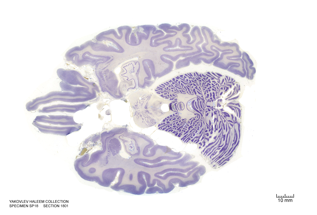
Figure 4.43: Horizontal section from The Human Brain Atlas at the Michigan State University Brain Biodiveristy Bank which acknowledges their support from the National Science Foundation.
In Figure 4.44, label the following structures:
- The 3d ventricle
- The optic nerve
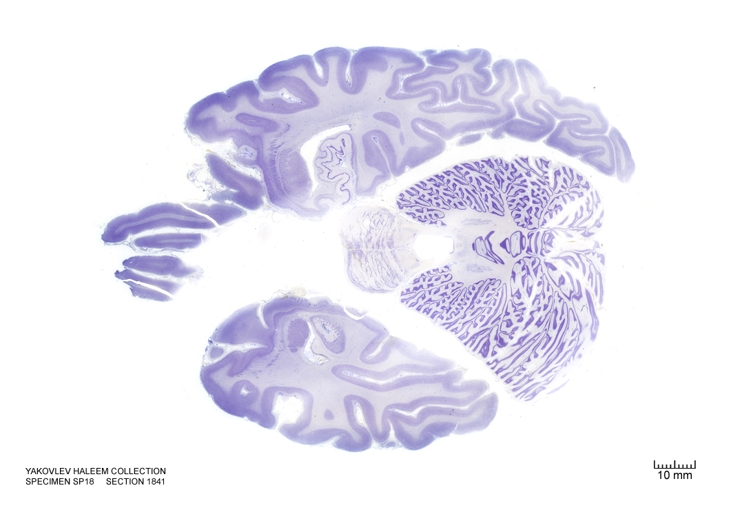
Figure 4.44: Horizontal section from The Human Brain Atlas at the Michigan State University Brain Biodiveristy Bank which acknowledges their support from the National Science Foundation.
In Figure 4.45, label the following structures:
- The 3d ventricle
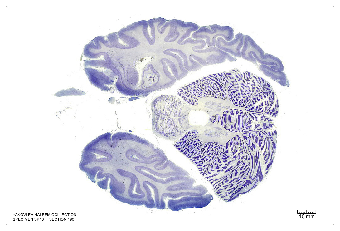
Figure 4.45: Horizontal section from The Human Brain Atlas at the Michigan State University Brain Biodiveristy Bank which acknowledges their support from the National Science Foundation.
In Figure 4.46, label the following structures:
- The 3d ventricle
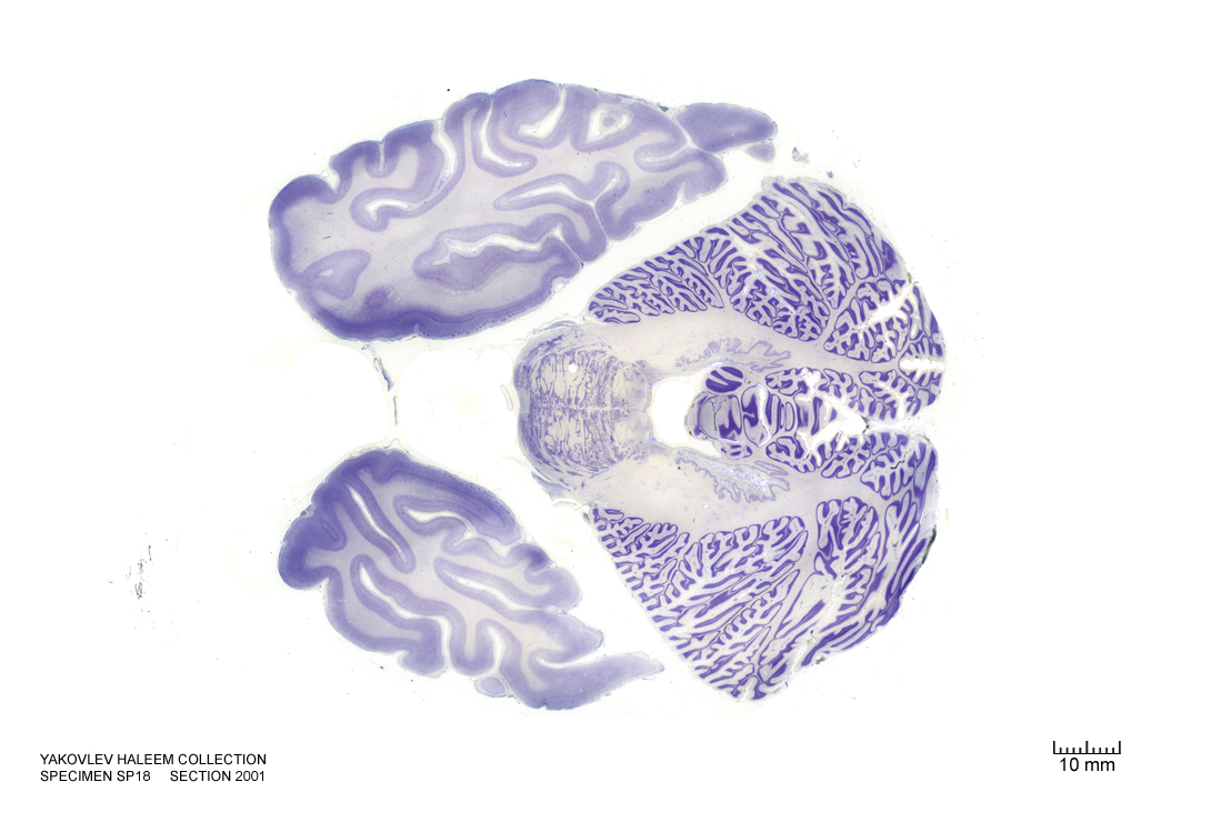
Figure 4.46: Horizontal section from The Human Brain Atlas at the Michigan State University Brain Biodiveristy Bank which acknowledges their support from the National Science Foundation.