9 Nematoda and Arthropoda
9.1 Nematodes
The nematodes or roundworms constitute the phylum Nematoda (also called Nemathelminthes). They are a diverse animal phylum inhabiting a broad range of environments. Nematode species can be difficult to distinguish, and although over 25,000 have been described, of which more than half are parasitic, it is estimated that over 40,000 species exist. Nematodes are classified along with insects and other moulting animals in the clade Ecdysozoa, and, unlike flatworms, have tubular digestive systems with openings at both ends. A characteristic of Nematoda is the one-way digestive tract, with a pseudocoelom (body cavity made up of only an ectoderm and endoderm).
Nematodes have successfully adapted to nearly every ecosystem from marine (salt water) to fresh water, to soils, and from the polar regions to the tropics, as well as the highest to the lowest of elevations. They are ubiquitous in freshwater, marine, and terrestrial environments, where they often outnumber other animals in both individual and species counts, and are found in locations as diverse as mountains, deserts and oceanic trenches. They are found in every part of the earth’s lithosphere, even at great depths, 0.9–3.6 km, below the surface of the Earth in gold mines in South Africa. They represent 90% of all animals on the ocean floor. Their numerical dominance, often exceeding a million individuals per square meter and accounting for about 80% of all individual animals on earth, their diversity of life cycles, and their presence at various trophic levels point at an important role in many ecosystems. The many parasitic forms include pathogens in most plants and animals. A third of the genera occur as parasites of vertebrates; about 35 nematode species occur in humans.
Nematodes are small, slender worms: typically approximately 5 to 100 µm thick, and 0.1 to 2.5 mm long. The smallest nematodes are microscopic, while free-living species can reach as much as 5 cm, and some parasitic species are larger still, reaching over 1 m in length. The body is often ornamented with ridges, rings, bristles, or other distinctive structures.
The head of a nematode is relatively distinct. Whereas the rest of the body is bilaterally symmetrical, the head is radially symmetrical, with sensory bristles and, in many cases, solid ‘head-shields’ radiating outwards around the mouth. The mouth has either three or six lips, which often bear a series of teeth on their inner edges. An adhesive ‘caudal gland’ is often found at the tip of the tail.
The epidermis is either a syncytium or a single layer of cells, and is covered by a thick collagenous cuticle. The cuticle is often of complex structure, and may have two or three distinct layers. Underneath the epidermis lies a layer of longitudinal muscle cells. The relatively rigid cuticle works with the muscles to create a hydroskeleton as nematodes lack circumferential muscles. Projections run from the inner surface of muscle cells towards the nerve cords; this is a unique arrangement in the animal kingdom, in which nerve cells normally extend fibres into the muscles rather than vice versa.
The oral cavity is lined with cuticle, which is often strengthened with ridges or other structures, and, especially in carnivorous species, may bear a number of teeth. The mouth often includes a sharp stylet, which the animal can thrust into its prey. In some species, the stylet is hollow, and can be used to suck liquids from plants or animals. The oral cavity opens into a muscular, sucking pharynx, also lined with cuticle. Digestive glands are found in this region of the gut, producing enzymes that start to break down the food. In stylet-bearing species, these may even be injected into the prey. There is no stomach, with the pharynx connecting directly to a intestine (not surrounded by smooth muscle) that forms the main length of the gut. This produces further enzymes, and also absorbs nutrients through its single cell thick lining. The last portion of the intestine is lined by cuticle, forming a rectum, which expels waste through the anus just below and in front of the tip of the tail. Movement of food through the digestive system is the result of body movements of the worm. The intestine has valves or sphincters at either end to help control the movement of food through the body.
Nitrogenous waste is excreted in the form of ammonia through the body wall, and is not associated with any specific organs. However, the structures for excreting salt to maintain osmoregulation are typically more complex. In many marine nematodes, one or two unicellular ‘renette glands’ excrete salt through a pore on the underside of the animal, close to the pharynx. In most other nematodes, these specialized cells have been replaced by an organ consisting of two parallel ducts connected by a single transverse duct. This transverse duct opens into a common canal that runs to the excretory pore.
Four peripheral nerves run the length of the body on the dorsal, ventral, and lateral surfaces. Each nerve lies within a cord of connective tissue lying beneath the cuticle and between the muscle cells. The ventral nerve is the largest, and has a double structure forward of the excretory pore. The dorsal nerve is responsible for motor control, while the lateral nerves are sensory, and the ventral combines both functions. The nervous system is also the only place in the nematode body that contains cilia, which are all non-motile and with a sensory function. At the anterior end of the animal, the nerves branch from a dense, circular nerve (nerve ring) round surrounding the pharynx, and serving as the brain. Smaller nerves run forward from the ring to supply the sensory organs of the head. The bodies of nematodes are covered in numerous sensory bristles and papillae that together provide a sense of touch. Behind the sensory bristles on the head lie two small pits, or ‘amphids’. These are well supplied with nerve cells, and are probably chemoreception organs. A few aquatic nematodes possess what appear to be pigmented eye-spots, but is unclear whether or not these are actually sensory in nature.
Most nematode species are dioecious, with separate male and female individuals, though some, such as Caenorhabditis elegans, are androdioecious, consisting of hermaphrodites and rare males. Both sexes possess one or two tubular gonads. In males, the sperm are produced at the end of the gonad and migrate along its length as they mature. The testis opens into a relatively wide seminal vesicle and then during intercourse into a glandular and muscular ejaculatory duct associated with the vas deferens and cloaca. In females, the ovaries each open into an oviduct (in hermaphrodites, the eggs enter a spermatheca first) and then a glandular uterus. The uteri both open into a common vulva/ vagina, usually located in the middle of the morphologically ventral surface.
Reproduction is usually sexual, though hermaphrodites are capable of self-fertilization. Males are usually smaller than females/ hermaphrodites (often much smaller) and often have a characteristically bent or fan-shaped tail. During copulation, one or more chitinized spicules move out of the cloaca and are inserted into the genital pore of the female. Amoeboid sperm crawl along the spicule into the female worm.
Eggs may be embryonated (containing an embryo) or unembryonated when passed by the female, meaning their fertilized eggs may not yet be developed. A few species are known to be ovoviviparous. The eggs are protected by an outer shell, secreted by the uterus. In free-living roundworms, the eggs hatch into larvae, which appear essentially identical to the adults, except for an underdeveloped reproductive system; in parasitic roundworms, the life cycle is often much more complicated.
Nematodes as a whole possess a wide range of modes of reproduction. Some nematodes, such as Heterorhabditis spp., undergo a process called endotokia matricida: intrauterine birth causing maternal death. Some nematodes are hermaphroditic, and keep their self-fertilized eggs inside the uterus until they hatch. The juvenile nematodes will then ingest the parent nematode. This process is significantly promoted in environments with a low food supply.
Nematodes commonly parasitic on humans include ascarids (Ascaris), filarias, hookworms, pinworms (Enterobius), and whipworms (Trichuris trichiura). The species Trichinella spiralis, commonly known as the ‘trichina worm’, occurs in rats, pigs, and humans, and is responsible for the disease trichinosis.
9.2 Ascaris
Ascaris is a genus of parasitic nematode worms known as the “small intestinal roundworms”. One species, Ascaris lumbricoides, affects humans and causes the disease ascariasis. Their eggs are deposited in feces and soil. Plants with the eggs on them infect any or ganism that consumes them. A. lumbricoides is the largest intestinal roundworm and is the most common helminth infection of humans worldwide. Infestation can cause morbidity by compromising nutritional status, affecting cognitive processes, inducing tissue reactions such as granuloma to larval stages, and by causing intestinal obstruction, which can be fatal. The body is long, cylindrical, fusiform (pointed at both the ends), body wall is composed of cuticle, epidermis and musculature. Presence of a false body pseudocoelom not lined by epithelium. Digestive system is complete. Respiration by simple diffusion. Nervous system consists of a nerve ring and many longitudinal nerve cords. Only sexual reproduction. Sexes are separate with sexual dimorphism. Males are usually shorter than females.
9.3 View Prepared Slides of Ascaris
- Female Ascaris x.s. (Figure 9.1)
- Identify: oviduct, pseudocoelom, intestine with lumen (GVC), uterus, eggs, ventral nerve cord, longitudinal muscles, cuticle, excretory canal
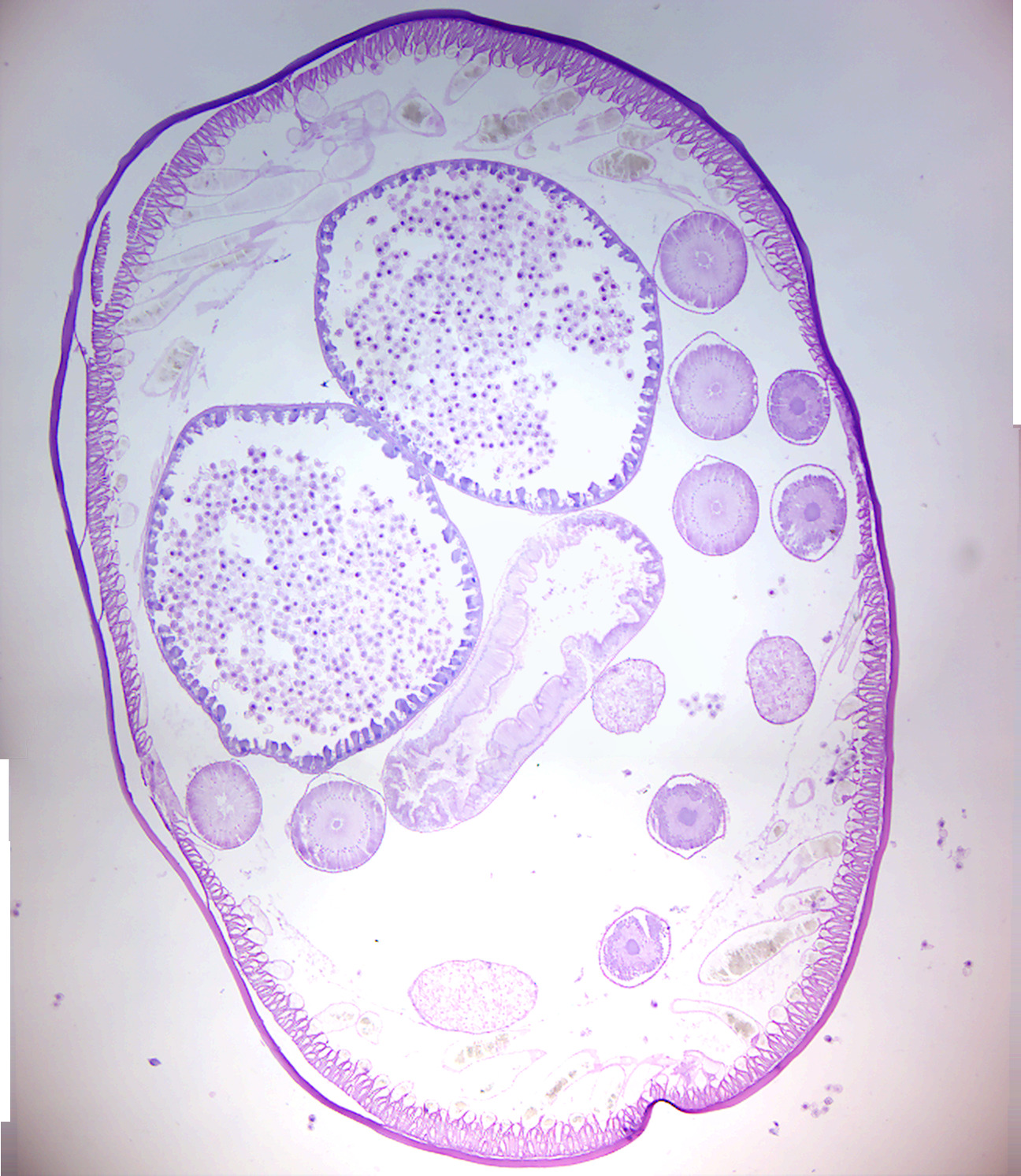
Figure 9.1: Female Ascaris.
9.4 Trichinella
Trichinella is the genus of parasitic roundworms of the phylum Nematoda that cause trichinosis (also known as trichinellosis). Members of this genus are often called trichinella or trichina worms. The genus was first recognised in a larval form in 1835. The L1 larvae live in a modified skeletal muscle cell. The adult worms occupy a membrane-bound portion of columnar epithelium, living as intracellular parasites. Infections with this genus have been reported from more than 150 different naturally or experimentally infected hosts. It has been shown to have a worldwide distribution in domestic and/or sylvatic animals. Trichinella is known as the smallest human nematode parasite, yet it is also the largest of all intracellular parasites. Oral ingestion of larvae-contaminated tissue is the usual route of infection. Cooking pork meat properly or by freezing pork, Trichinella infection can be prevented. However, freezing pork is not an effective method for killing larvae.
9.5 View Prepared Slides of Trichinella
- Trichinella encysted in muscle (Figure 9.2)
- Identify: muscle, cyst, larva
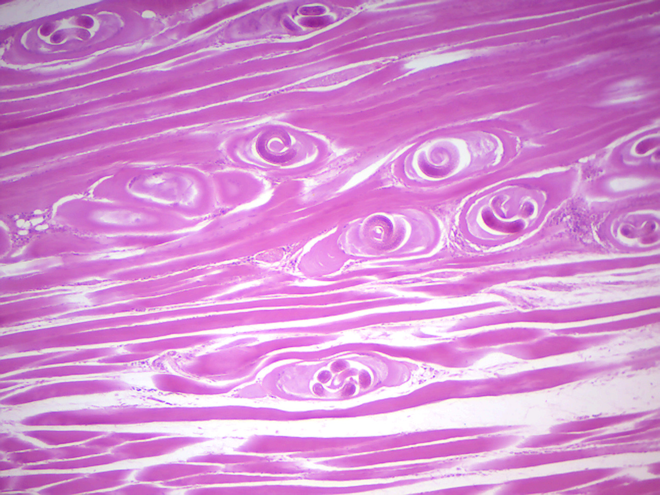
Figure 9.2: Trichinella encysted in muscle.
9.6 Vinegar eels
Turbatrix aceti (vinegar eels, vinegar nematode) are free-living nematodes that feed on the microbial culture, called mother of vinegar used to create vinegar, and may be found in unfiltered vinegar. Vinegar eels are often given to fry (baby fish) as a live food, like microworms. Although they are harmless and non-parasitic, leaving eels in vinegar is considered objectionable in the United States and is not permitted in vinegar destined for American consumers. Manufacturers normally filter and pasteurize their product prior to bottling, destroying the live bacterial and yeast culture that these nematodes require for sustenance.
9.7 Arthropods
Arthropods (from arthron, “joint” and pous, “foot”) are invertebrate animals having an exoskeleton (external skeleton), a segmented body, and paired jointed appendages. Arthropods form the phylum Euarthropoda, which includes insects, arachnids, myriapods, and crustaceans. Arthropods are characterized by their jointed limbs and cuticle made of chitin, often mineralized with calcium carbonate. The arthropod body plan consists of segments, each with a pair of appendages. The rigid cuticle inhibits growth, so arthropods replace it periodically by molting.
Their versatility has enabled them to become the most species-rich members of all ecological guilds in most environments. They have over a million described species, making up more than 80% of all described living animal species, some of which, unlike most animals, are very successful in dry environments.
Arthropods range in size from the microscopic crustacean Stygotantulus up to the Japanese spider crab. Arthropods’ primary internal cavity is a hemocoel, which accommodates their internal organs, and through which their hemolymph – analogue of blood – circulates; they have open circulatory systems. Like their exteriors, the internal organs of arthropods are generally built of repeated segments. Their nervous system is “ladder-like”, with paired ventral nerve cords running through all segments and forming paired ganglia in each segment.
Their heads are formed by fusion of varying numbers of segments, and their brains are formed by fusion of the ganglia of these segments and encircle the esophagus. The respiratory and excretory systems of arthropods vary, depending as much on their environment as on the subphylum to which they belong.
Their vision relies on various combinations of compound eyes and pigment-pit ocelli: in most species, the ocelli can only detect the direction from which light is coming, and the compound eyes are the main source of information, but the main eyes of spiders are ocelli that can form images and, in a few cases, can swivel to track prey. Arthropods also have a wide range of chemical and mechanical sensors, mostly based on modifications of the many setae (bristles) that project through their cuticles. Arthropods’ methods of reproduction and development are diverse; all terrestrial species use internal fertilization, but this is often by indirect transfer of the sperm via an appendage or the ground, rather than by direct injection.
Aquatic species use either internal or external fertilization. Almost all arthropods lay eggs, but scorpions give birth to live young after the eggs have hatched inside the mother. Arthropod hatchlings vary from miniature adults to grubs and caterpillars that lack jointed limbs and eventually undergo a total metamorphosis to produce the adult form. The level of maternal care for hatchlings varies from nonexistent to the prolonged care provided by scorpions.
The evolutionary ancestry of arthropods dates back to the Cambrian period. The group is generally regarded as monophyletic, and many analyses support the placement of arthropods with cycloneuralians (or their constituent clades) in a superphylum Ecdysozoa.
Arthropods contribute to the human food supply both directly as food, and more importantly indirectly as pollinators of crops. Some species are known to spread severe disease to humans, livestock, and crops.
Arthropods belong to the phylum Euarthropoda. The phylum is sometimes called Arthropoda, but strictly this term denotes a (putative - see Tactopoda) clade that also encompasses Phylum Onychophora.
The appendages of arthropods may be either biramous or uniramous. A uniramous limb comprises a single series of segments attached end-to-end. A biramous limb, however, branches into two, and each branch consists of a series of segments attached end-to-end.
The phylum Euarthropoda is typically subdivided into five subphyla, of which one is extinct:
- Trilobites are a group of formerly numerous marine animals that disappeared in the Permian–Triassic extinction event, though they were in decline prior to this killing blow, having been reduced to one order in the Late Devonian extinction.
- Chelicerates include horseshoe crabs, spiders, mites, scorpions and related organisms. They are characterized by the presence of chelicerae, appendages just above / in front of the mouth. Chelicerae appear in scorpions and horseshoe crabs as tiny claws that they use in feeding, but those of spiders have developed as fangs that inject venom.
- Myriapods comprise millipedes, centipedes, and their relatives and have many body segments, each segment bearing one or two pairs of legs (or in a few cases being legless). They are sometimes grouped with the hexapods.
- Crustaceans are primarily aquatic (a notable exception being woodlice) and are characterized by having biramous appendages. They include lobsters, crabs, barnacles, crayfish, shrimp and many others.
- Hexapods comprise insects and three small orders of insect-like animals with six thoracic legs. They are sometimes grouped with the myriapods, in a group called Uniramia, though genetic evidence tends to support a closer relationship between hexapods and crustaceans.
9.8 Crayfish
Crayfish, also known as crawfish, crawdads, freshwater lobsters, mountain lobsters, mudbugs or yabbies, are freshwater crustaceans resembling small lobsters, to which they are related. They breathe through feather-like gills. Some species are found in brooks and streams where there is running fresh water, while others thrive in swamps, ditches, and paddy fields. Crayfish feed on animals and plants, either living or decomposing, and detritus. Its body is made up of twenty body segments grouped into two main body parts, the cephalothorax and the abdomen. Each segment may possess one pair of appendages, although in various groups these may be reduced or missing. On average, crayfish grow to 17.5 centimeters in length, but some grow larger. Walking legs have a small claw at the end.
Please watch the following video showing the external anatomy of a crayfish:
Please watch the following video showing the internal anatomy of a crayfish:
9.9 Crayfish Dissection
- Get a dissction pan, scissorc, forceps, and a pointer.
- Get a cray fish.
- Place a crayfish on its side in a dissection tray (Fig. 9.3.
- Locate the cephalothorax and the abdomen. The carapace, a shield of chitin, covers the dorsal surface of the cephalothorax. On the carapace, observe an indentation, the cervical groove, that extends across the midregion and separates the head and thoracic regions. On the thoracic region, locate the prominent suture or indentation on the cephalothorax that defines a central area separate from the sides. Note the individual segments of the abdomen.
- Turn the crayfish with its dorsal side upward.
- Locate the rostrum, which is the pointed extension of the carapace at the head of the animal. Locate the two eyes at the end of stalks.
- Locate the five pairs of appendages on the head region. First locate the antennules in the most anterior segment. Behind them observe the much longer pair of antennae.
- Turn the crayfish around with its ventral side upward.
- Locate the mouth. Observe the mandibles (jaws), behind the antennae.
- Locate the two pairs of maxillae, which are the last appendages in the cephalic region.
- On the thoracic portion of the cephalothorax, observe the three pointed maxillipeds.
- On the abdomen, observe six distinct segments. Observe a pair of swimmerets on each of the first five segments.
- On the last abdominal segment, observe a pair of pointed appendages modified into a pair of uropods. In the middle of the uropods, locate the triangular-shaped telson.
- Determine the sex of your specimen. Males (Fig. 9.4) have hardened gonapods and hooks on the third pair of legs. Females (9.6) have an opening to a seminal receptacle between the fifth pair of legs.
- Turn the crayfish around so that its dorsal side is upward.
- Use one hand to hold the crayfish dorsal side up in the dissecting tray, use scissors to carefully cut through the back of the carapace along the dotted line shown in figure 9.8 below. Cut along the indentations that separate the thoracic portion of the carapace into three regions. Start the cut at the posterior edges of the carapace, and extend it along both sides in the cephalic region.
- Using the forceps, carefully and slowly lift away the carapace.
- Place the crayfish on its side (head facing left). Using scissors, cut starting at the base along the dotted line shown in Figure 9.9. Extend the cut line forward toward the rostrum (at the top of the head).
- Use forceps to carefully lift away the remaining parts of the carapace, exposing the underlying gills and other organs.
- Locate and identify the organs of the digestive and excretory systems. Locate the maxillae that pass the pieces of food into the mouth. The food travels down the short esophagus into the lobed (two-part) stomach (Fig. 9.10). Locate the digestive gland, which produces digestive substances and from which the absorption of nutrients occurs. Undigested material passes into the intestine. Observe that the intestine is attached to the stomach. The undigested material is eliminated from the anus. Locate the green glands at the base of the antennae of the maxilla (Fig. 9.13). The green glands are part of the excretory system and eliminate toxic and waste products similar to the function of the kidneys in mammals.
- Locate the gills, which are feather-like structures found underneath the carapace and attached to the chelipeds and walking legs.
- Locate the dorsal tubular heart and several arteries. Notice the holes in the heart (Fig. 9.11) indicating that the crayfish has an open circulatory system in which the blood flows from arteries into sinuses, or spaces, in tissues. The blood flows over the gills before returning to the heart through the holes.
- Find the ventral nerve cord. Locate a ganglion, one of the enlargements of the ventral nerve cord. Locate the dorsal brain (Fig. 9.10) which is located just behind the compound eyes. Note the two large nerves that lead from the brain, around the esophagus, and join the ventral nerve cord.
- Locate and identify the organs of the reproductive system. If the specimen is male, locate the testis. The testis is the long, white organ under the heart. If the specimen is a female, locate the bi-lobed ovary. It is in the same relative position as the testis, but the ovary appears as a large, reddish mass under the heart.

Figure 9.3: Crayfish

Figure 9.4: Ventral view of a male crayfish. Notice the gonapods and hooks.
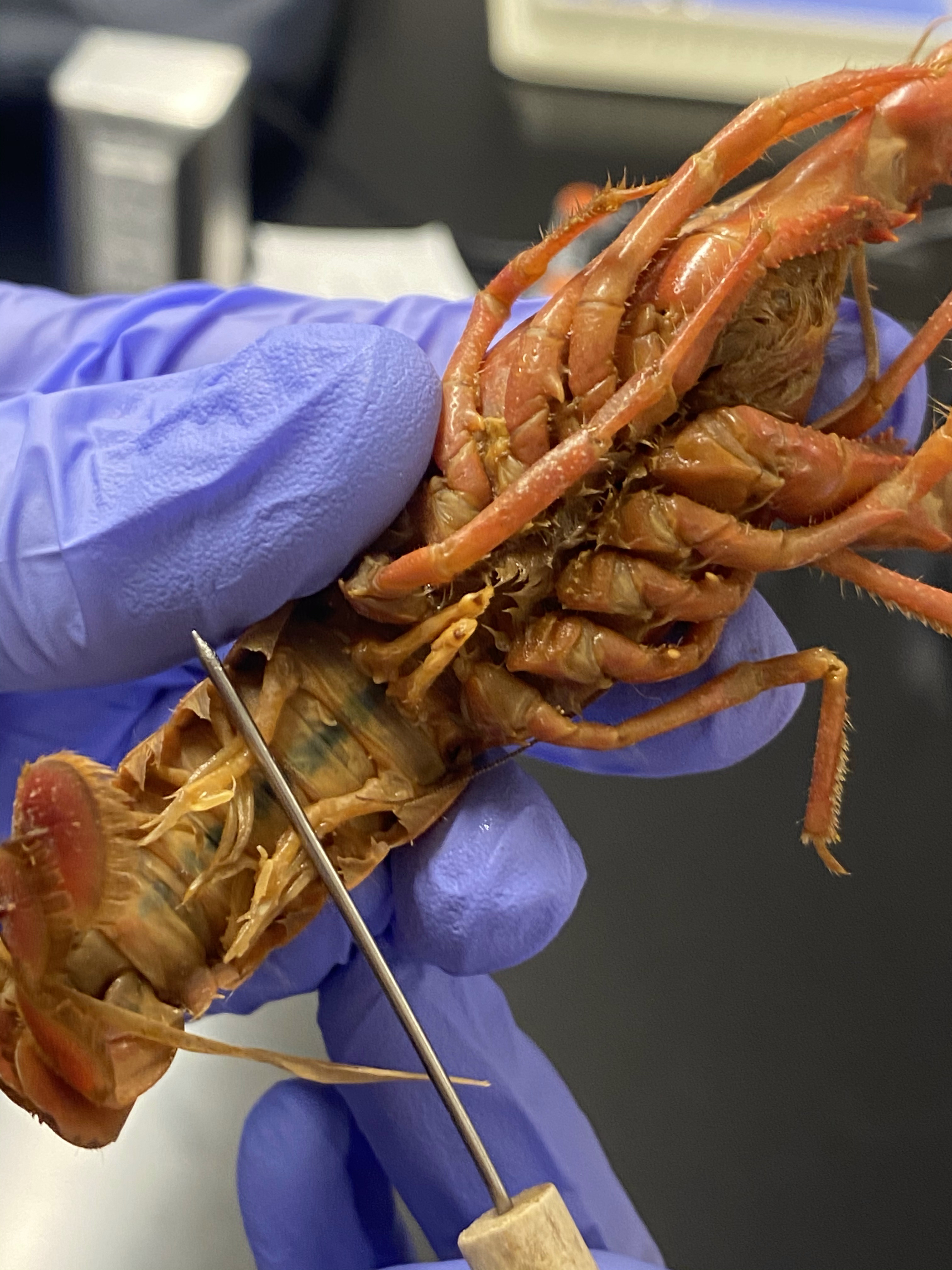
Figure 9.5: Ventral view of a male crayfish.

Figure 9.6: Ventral view of a female crayfish. Notice the central opening to the seminal receptacle.
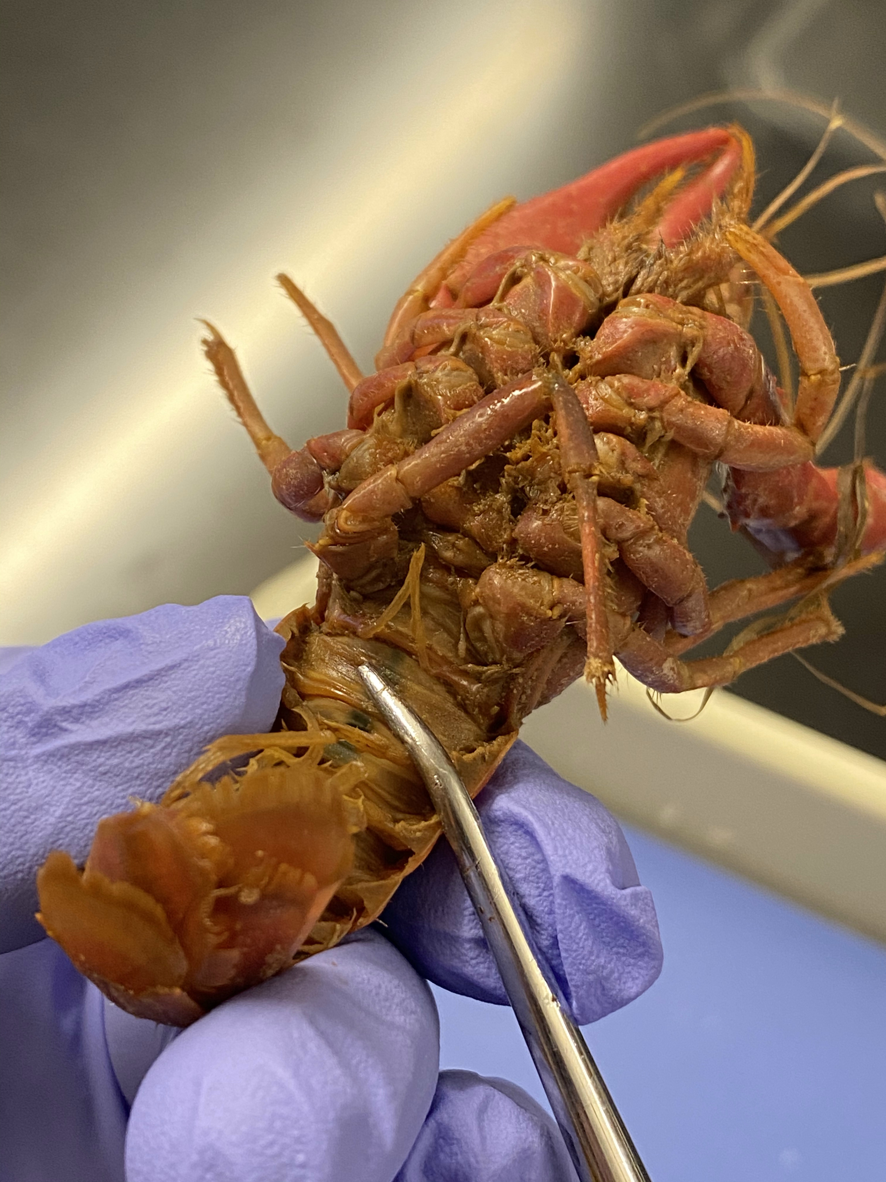
Figure 9.7: Ventral view of a female crayfish.

Figure 9.8: Dorsal view of the crayfish. Cut along the dotted lines.

Figure 9.9: Lateral view of the crayfish. Cut along the dotted lines.
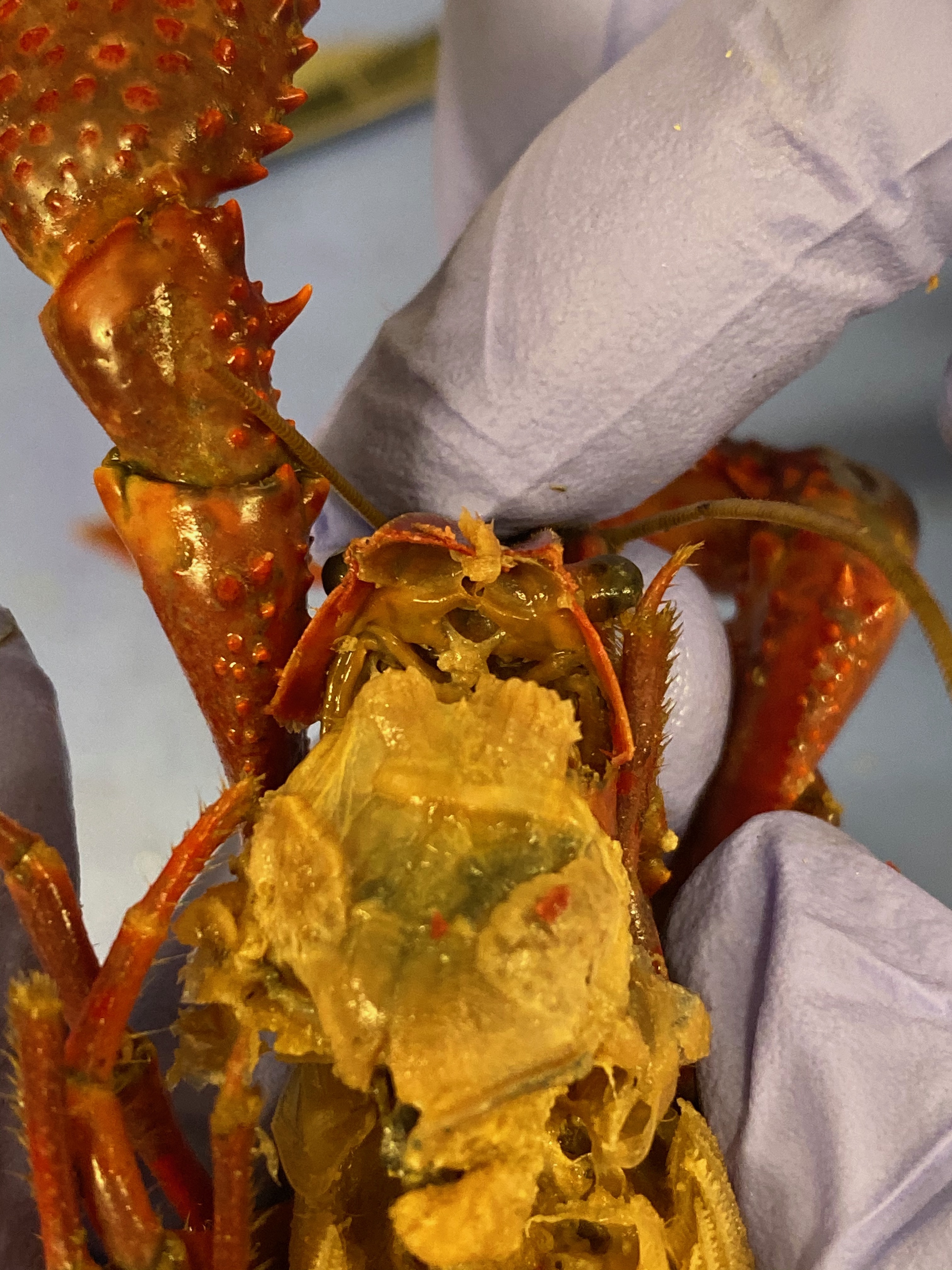
Figure 9.10: The crayfish brain and cardiac and pyloric stomach.
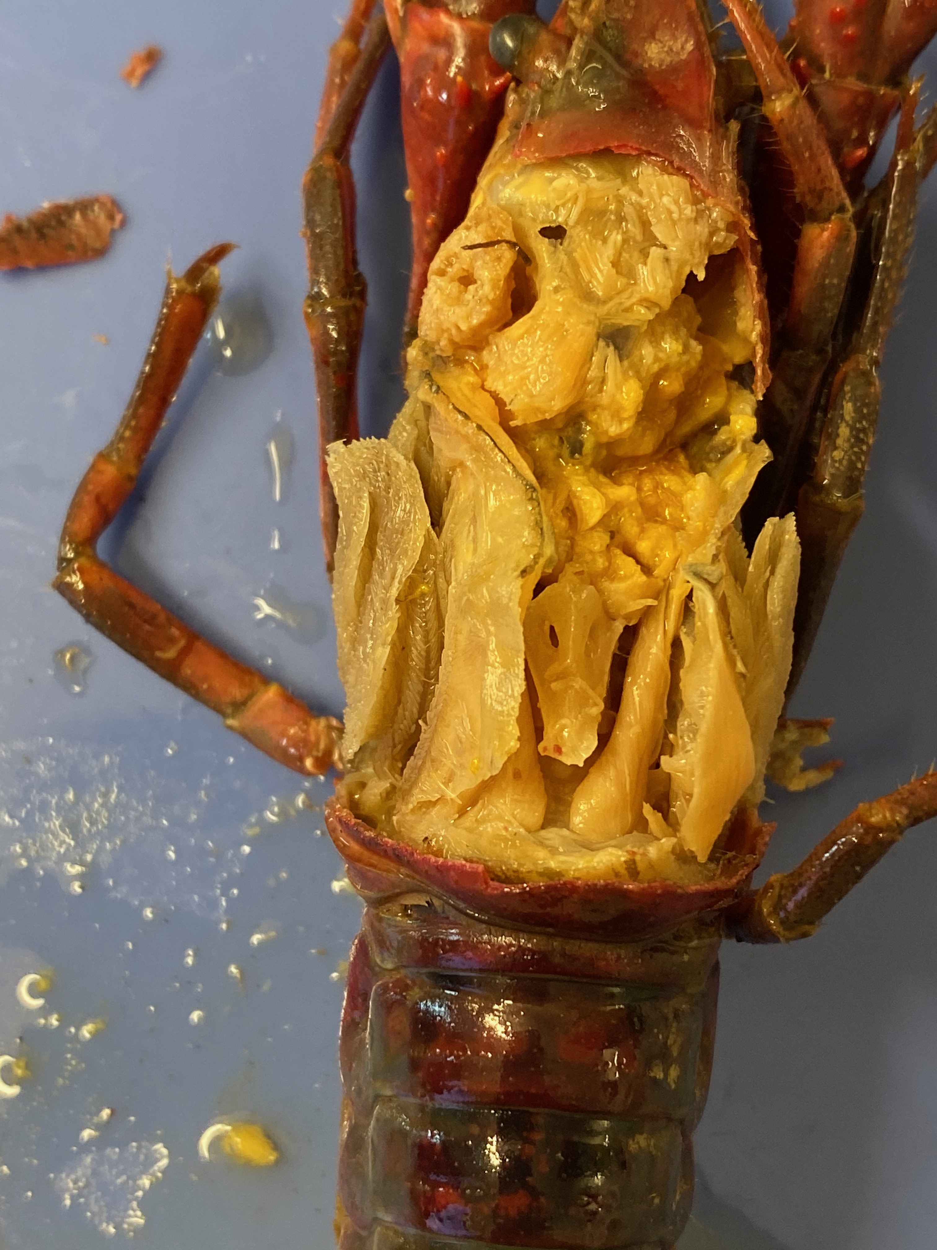
Figure 9.11: The crayfish heart. Notice the holes in the lateral walls.
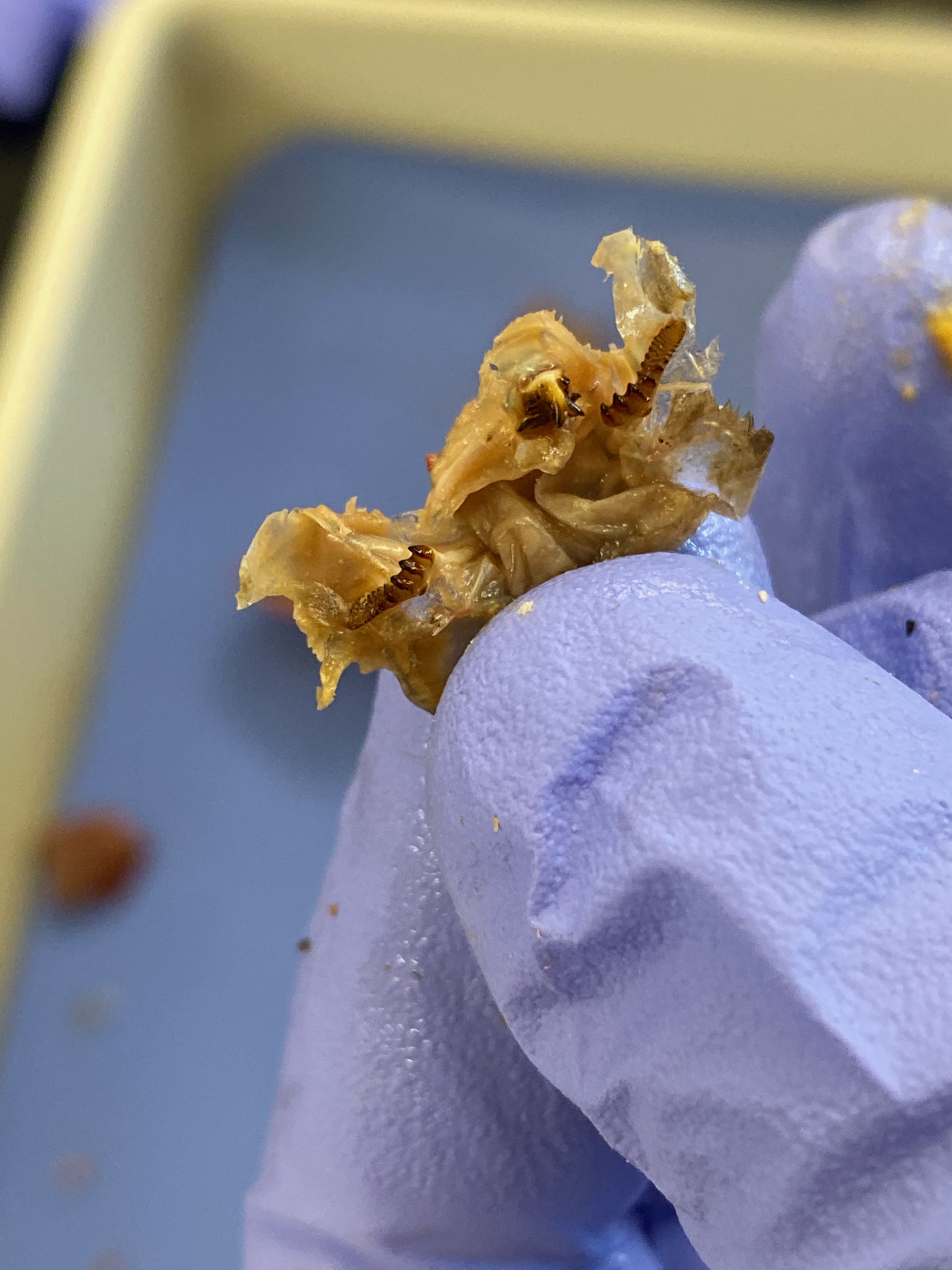
Figure 9.12: Teeth in the stomach of the crayfish.
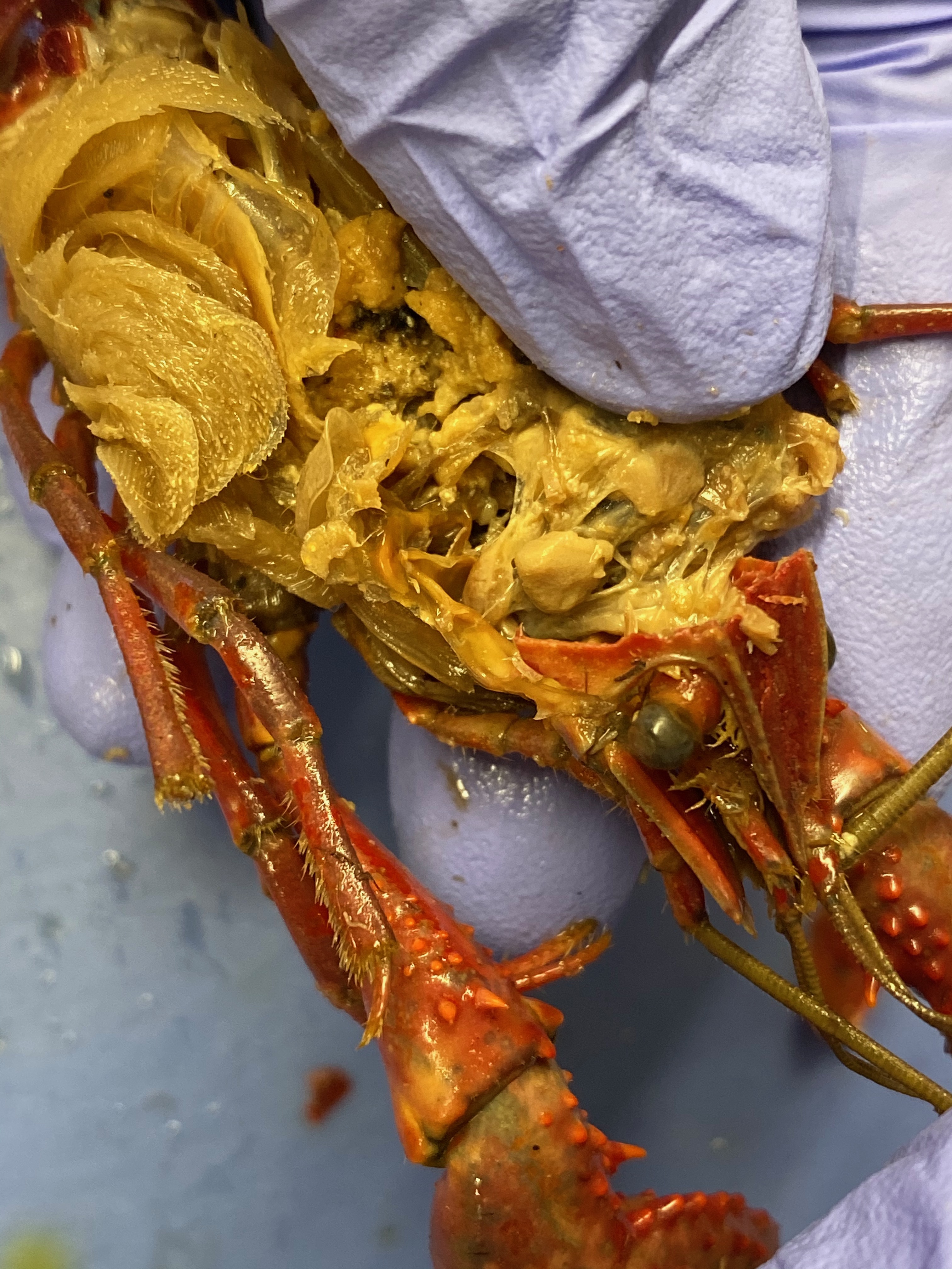
Figure 9.13: The green glands (also called maxillary or antennal glands) of the crayfish.
9.10 Cleaning up
- Dispose of the remains of the crayfish in the red biohazard bins.
- Clean the dissection tray and instruments and return them to the place where you picked them up.
- Clean table tops with red bottled sanitizer.
- Wash your hands.
9.11 View Living Organisms
- Vinegar eels
- Mixed crustaceans
- Look for isopods (sow bugs), amphipods, copepods, water fleas (Daphnia), ostracods, and fairy shrimp.
9.12 Review Questions
- What are nematodes?
- What are arthropods?
- What is an exoskeleton?
- What are crustaceans?